

Transplantation of neural precursors generated from spinal progenitor cells reduces inflammation in spinal cord injury via NF-κB pathway inhibition
- PMID: 30654804
- PMCID: PMC6335809
- DOI: 10.1186/s12974-019-1394-7
Transplantation of neural precursors generated from spinal progenitor cells reduces inflammation in spinal cord injury via NF-κB pathway inhibition
Abstract
Background: Traumatic spinal cord injury (SCI) triggers a chain of events that is accompanied by an inflammatory reaction leading to necrotic cell death at the core of the injury site, which is restricted by astrogliosis and apoptotic cell death in the surrounding areas. Activation of nuclear factor-κB (NF-κB) signaling pathway has been shown to be associated with inflammatory response induced by SCI. Here, we elucidate the pattern of activation of NF-κB in the pathology of SCI in rats and investigate the effect of transplantation of spinal neural precursors (SPC-01) on its activity and related astrogliosis.
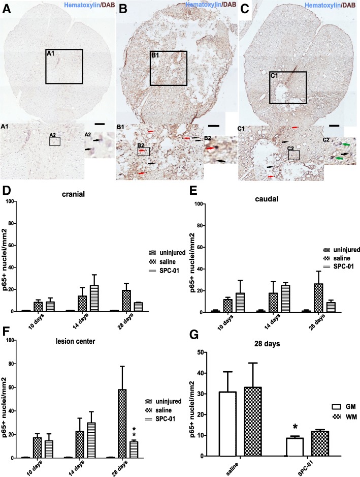
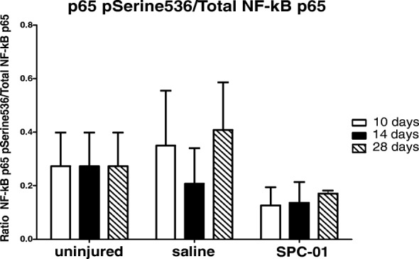
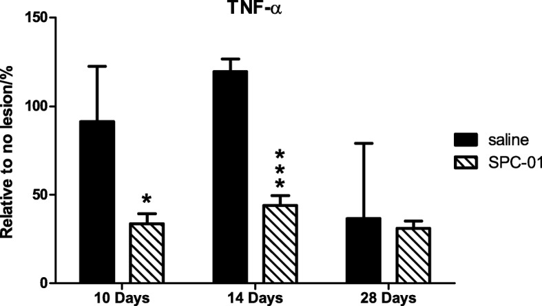
Methods: Using a rat compression model of SCI, we transplanted SPC-01 cells or injected saline into the lesion 7 days after SCI induction. Paraffin-embedded sections were used to assess p65 NF-κB nuclear translocation at days 1, 3, 7, 10, 14, and 28 and to determine levels of glial scaring, white and gray matter preservation, and cavity size at day 28 after SCI. Additionally, levels of p65 phosphorylated at Serine536 were determined 10, 14, and 28 days after SCI as well as levels of locally secreted TNF-α.
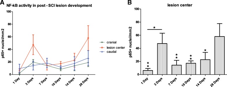
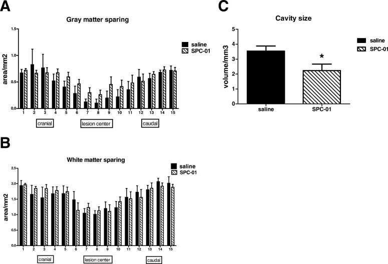
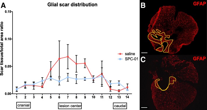
Results: We determined a bimodal activation pattern of canonical p65 NF-κB signaling pathway in the pathology of SCI with peaks at 3 and 28 days after injury induction. Transplantation of SCI-01 cells resulted in significant downregulation of TNF-α production at 10 and 14 days after SCI and in strong inhibition of p65 NF-κB activity at 28 days after SCI, mainly in the gray matter. Moreover, reduced formation of glial scar was found in SPC-01-transplanted rats along with enhanced gray matter preservation and reduced cavity size.
Conclusions: The results of this study demonstrate strong immunomodulatory properties of SPC-01 cells based on inhibition of a major signaling pathway. Canonical NF-κB pathway activation underlines much of the immune response after SCI including cytokine, chemokine, and apoptosis-related factor production as well as immune cell activation and infiltration. Reduced inflammation may have led to observed tissue sparing. Additionally, such immune response modulation could have impacted astrocyte activation resulting in a reduced glial scar.
Keywords: Inflammation; NF-κB; Spinal cord injury; Stem cells transplantation; TNF-α; p65.
Conflict of interest statement
Ethics approval and consent to participate
All experiments were performed in accordance with the European Communities Council Directive of 22nd of September 2010 (2010/63/EU) regarding the use of animals in research, and were approved by the Ethics Committee of the Institute of Experimental Medicine ASCR, Prague, Czech Republic under no: 277/2011 and 55/2017.
Consent for publication
Not applicable.
Competing interests
The authors declare that they have no competing interests.
Publisher’s Note
Springer Nature remains neutral with regard to jurisdictional claims in published maps and institutional affiliations.
Figures
Similar articles
-
Valproic acid attenuates traumatic spinal cord injury-induced inflammation via STAT1 and NF-κB pathway dependent of HDAC3.J Neuroinflammation. 2018 May 18;15(1):150. doi: 10.1186/s12974-018-1193-6. J Neuroinflammation. 2018. PMID: 29776446 Free PMC article.
-
A Comparative Study of Three Different Types of Stem Cells for Treatment of Rat Spinal Cord Injury.Cell Transplant. 2017 Apr 13;26(4):585-603. doi: 10.3727/096368916X693671. Epub 2016 Nov 2. Cell Transplant. 2017. PMID: 27938489 Free PMC article.
-
Effects of dihydroxylphenyl lactic acid on inflammatory responses in spinal cord injury.Brain Res. 2011 Feb 4;1372:160-8. doi: 10.1016/j.brainres.2010.11.089. Epub 2010 Dec 4. Brain Res. 2011. PMID: 21134362
-
The NF-κB Pathway: a Focus on Inflammatory Responses in Spinal Cord Injury.Mol Neurobiol. 2023 Sep;60(9):5292-5308. doi: 10.1007/s12035-023-03411-x. Epub 2023 Jun 7. Mol Neurobiol. 2023. PMID: 37286724 Review.
-
Stem cell-based cell therapy for spinal cord injury.Cell Transplant. 2007;16(4):355-64. doi: 10.3727/000000007783464885. Cell Transplant. 2007. PMID: 17658126 Review.
Cited by
-
Filling the Gap: Neural Stem Cells as A Promising Therapy for Spinal Cord Injury.Pharmaceuticals (Basel). 2019 Apr 29;12(2):65. doi: 10.3390/ph12020065. Pharmaceuticals (Basel). 2019. PMID: 31035689 Free PMC article. Review.
-
Changes in transcriptome profiling during the acute/subacute phases of contusional spinal cord injury in rats.Ann Transl Med. 2020 Dec;8(24):1682. doi: 10.21037/atm-20-6519. Ann Transl Med. 2020. PMID: 33490194 Free PMC article.
-
A comprehensive transcriptional reference for severity and progression in spinal cord injury reveals novel translational biomarker genes.J Transl Med. 2025 Feb 4;23(1):160. doi: 10.1186/s12967-024-06009-6. J Transl Med. 2025. PMID: 39905473 Free PMC article.
-
Role of Nuclear Factor Kappa B (NF-κB) Signalling in Neurodegenerative Diseases: An Mechanistic Approach.Curr Neuropharmacol. 2020;18(10):918-935. doi: 10.2174/1570159X18666200207120949. Curr Neuropharmacol. 2020. PMID: 32031074 Free PMC article. Review.
-
CYP11A1-derived vitamin D3 products protect against UVB-induced inflammation and promote keratinocytes differentiation.Free Radic Biol Med. 2020 Aug 1;155:87-98. doi: 10.1016/j.freeradbiomed.2020.05.016. Epub 2020 May 22. Free Radic Biol Med. 2020. PMID: 32447000 Free PMC article.
References
-
- Brambilla R, Persaud T, Hu X, Karmally S, Shestopalov VI, Dvoriantchikova G, et al. Transgenic inhibition of astroglial NF-kappa B improves functional outcome in experimental autoimmune encephalomyelitis by suppressing chronic central nervous system inflammation. J Immunol. 2009;182:2628–2640. doi: 10.4049/jimmunol.0802954. - DOI - PMC - PubMed
MeSH terms
Substances
Grants and funding
LinkOut - more resources
Full Text Sources
Medical
Molecular Biology Databases
