

Repeated gestational exposure to diesel engine exhaust affects the fetal olfactory system and alters olfactory-based behavior in rabbit offspring
- PMID: 30654819
- PMCID: PMC6335688
- DOI: 10.1186/s12989-018-0288-7
Repeated gestational exposure to diesel engine exhaust affects the fetal olfactory system and alters olfactory-based behavior in rabbit offspring
Abstract
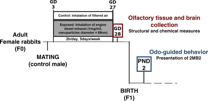
Background: Airborne pollution, especially from diesel exhaust (DE), is known to have a negative effect on the central nervous system in exposed human populations. However, the consequences of gestational exposure to DE on the fetal brain remain poorly explored, with various effects depending on the conditions of exposure, as well as little information on early developmental stages. We investigated the short-term effects of indirect DE exposure throughout gestation on the developing brain using a rabbit model. We analyzed fetal olfactory tissues at the end of gestation and tested behaviors relevant to pups' survival at birth. Pregnant dams were exposed by nose-only inhalation to either clean air or DE with a content of particles (DEP) adjusted to 1 mg/m3 by diluting engine exhaust, for 2 h/day, 5 days/week, from gestational day 3 (GD3) to day 27 (GD27). At GD28, fetal olfactory mucosa, olfactory bulbs and whole brains were collected for anatomical and neurochemical measurements. At postnatal day 2 (PND2), pups born from another group of exposed or control female were examined for their odor-guided behavior in response to the presentation of the rabbit mammary pheromone 2-methyl-3-butyn-2-ol (2MB2).
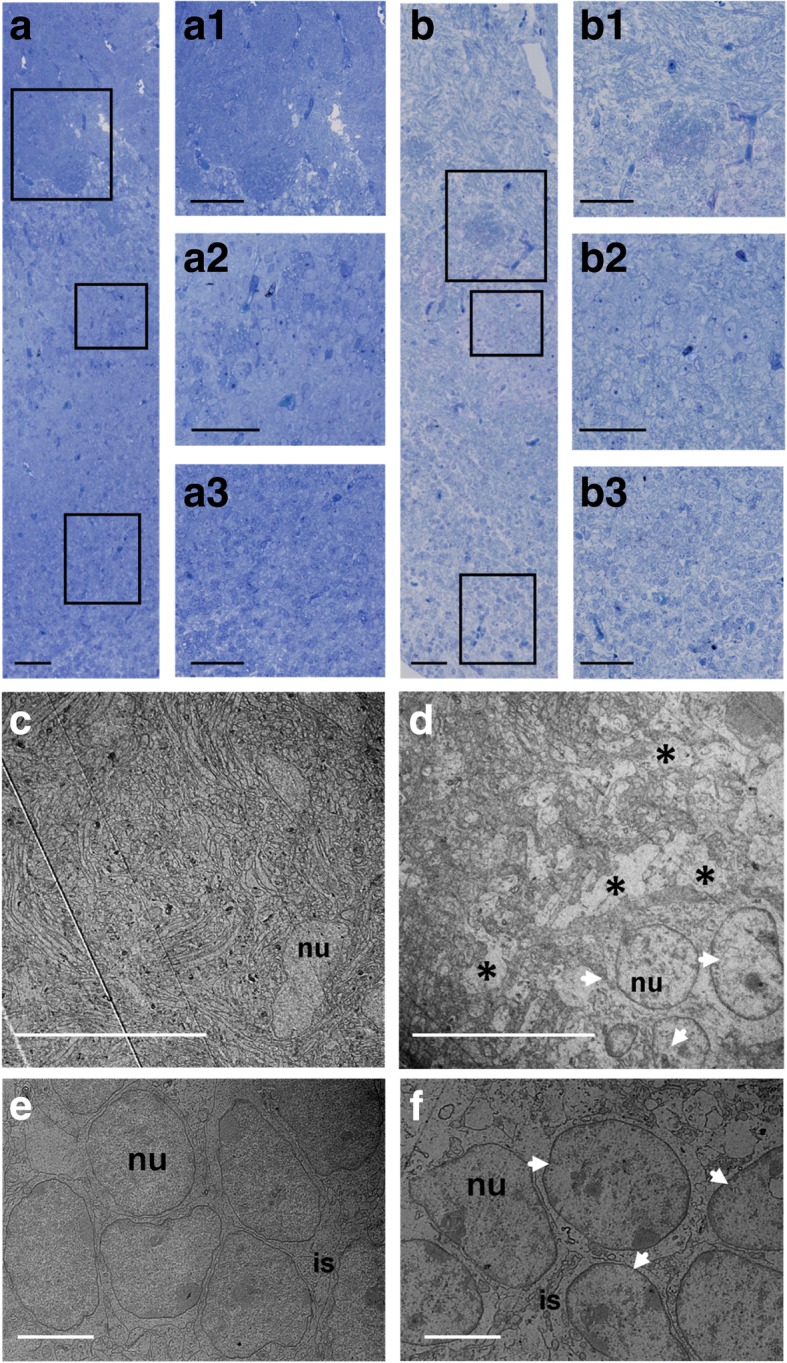
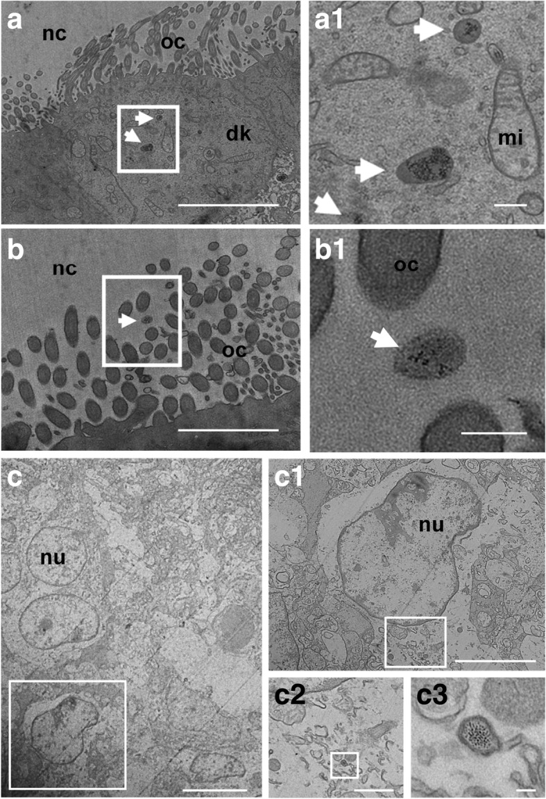
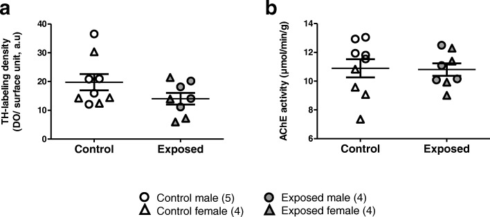
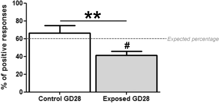
Results: At GD28, nano-sized particles were observed in cilia and cytoplasm of the olfactory sensory neurons in the olfactory mucosa and in the cytoplasm of periglomerular cells in the olfactory bulbs of exposed fetuses. Moreover, cellular and axonal hypertrophies were observed throughout olfactory tissues. Concomitantly, fetal serotoninergic and dopaminergic systems were affected in the olfactory bulbs. Moreover, the neuromodulatory homeostasis was disturbed in a sex-dependent manner in olfactory tissues. At birth, the olfactory sensitivity to 2MB2 was reduced in exposed PND2 pups.
Conclusion: Gestational exposure to DE alters olfactory tissues and affects monoaminergic neurotransmission in fetuses' olfactory bulbs, resulting in an alteration of olfactory-based behaviors at birth. Considering the anatomical and functional continuum between the olfactory system and other brain structures, and due to the importance of monoamine neurotransmission in the plasticity of neural circuits, such alterations could participate to disturbances in higher integrative structures, with possible long-term neurobehavioral consequences.
Keywords: Airborne pollution; Bulbar neurotransmitter disturbances; Diesel exhaust; Gestational exposure; Nano-particulate matter; Olfactory dysfunction; Olfactory toxicity; Olfactory-based behavior; Pheromonal response.
Conflict of interest statement
Ethics approval
Animals were treated according to the ethical standards defined by the National Institute for Agronomic Research (INRA) for animal health and care with strict compliance with the EEC recommendations (no. 86/609) and the 2010/63/EU directive on the protection of animals used for scientific purposes. The local ethical committee (N°45 in the French National register) approved the experimentation under N°12/102.
Consent for publication
Not applicable.
Competing interests
The authors declare that they have no competing interests.
Publisher’s Note
Springer Nature remains neutral with regard to jurisdictional claims in published maps and institutional affiliations.
Figures

Similar articles
-
Dopaminergic and serotonergic changes in rabbit fetal brain upon repeated gestational exposure to diesel engine exhaust.Arch Toxicol. 2021 Sep;95(9):3085-3099. doi: 10.1007/s00204-021-03110-3. Epub 2021 Jun 29. Arch Toxicol. 2021. PMID: 34189592
-
Placental-fetal distribution of carbon particles in a pregnant rabbit model after repeated exposure to diluted diesel engine exhaust.Part Fibre Toxicol. 2023 May 18;20(1):20. doi: 10.1186/s12989-023-00531-z. Part Fibre Toxicol. 2023. PMID: 37202804 Free PMC article.
-
In utero exposure to a low concentration of diesel exhaust affects spontaneous locomotor activity and monoaminergic system in male mice.Part Fibre Toxicol. 2010 Mar 23;7:7. doi: 10.1186/1743-8977-7-7. Part Fibre Toxicol. 2010. PMID: 20331848 Free PMC article.
-
Endocrine-disrupting activity of chemicals in diesel exhaust and diesel exhaust particles.Environ Sci. 2004;11(1):33-45. Environ Sci. 2004. PMID: 15746887 Review.
-
Developmental toxicity of diesel exhaust: a review of studies in experimental animals.Reprod Toxicol. 2013 Dec;42:1-17. doi: 10.1016/j.reprotox.2013.06.074. Epub 2013 Jul 4. Reprod Toxicol. 2013. PMID: 23831197 Review.
Cited by
-
Effects of intranasal instillation of nanoparticulate matter in the olfactory bulb.Sci Rep. 2021 Aug 20;11(1):16997. doi: 10.1038/s41598-021-96593-0. Sci Rep. 2021. PMID: 34417533 Free PMC article.
-
Effects of air pollution on the nervous system and its possible role in neurodevelopmental and neurodegenerative disorders.Pharmacol Ther. 2020 Jun;210:107523. doi: 10.1016/j.pharmthera.2020.107523. Epub 2020 Mar 9. Pharmacol Ther. 2020. PMID: 32165138 Free PMC article. Review.
-
Deciphering the Impact of Early-Life Exposures to Highly Variable Environmental Factors on Foetal and Child Health: Design of SEPAGES Couple-Child Cohort.Int J Environ Res Public Health. 2019 Oct 14;16(20):3888. doi: 10.3390/ijerph16203888. Int J Environ Res Public Health. 2019. PMID: 31615055 Free PMC article.
-
Label-free detection of uptake, accumulation, and translocation of diesel exhaust particles in ex vivo perfused human placenta.J Nanobiotechnology. 2021 May 17;19(1):144. doi: 10.1186/s12951-021-00886-5. J Nanobiotechnology. 2021. PMID: 34001140 Free PMC article.
-
Developmental impact of air pollution on brain function.Neurochem Int. 2019 Dec;131:104580. doi: 10.1016/j.neuint.2019.104580. Epub 2019 Oct 15. Neurochem Int. 2019. PMID: 31626830 Free PMC article. Review.
References
Publication types
MeSH terms
Substances
Grants and funding
LinkOut - more resources
Full Text Sources
Medical
