Review
. 2019 Jan 22;73(2):190-209.
doi: 10.1016/j.jacc.2018.09.089.

Endothelial to Mesenchymal Transition in Cardiovascular Disease: JACC State-of-the-Art Review

Affiliations
Review

Endothelial to Mesenchymal Transition in Cardiovascular Disease: JACC State-of-the-Art Review

Jason C Kovacic et al. J Am Coll Cardiol. .

Abstract

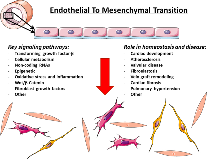
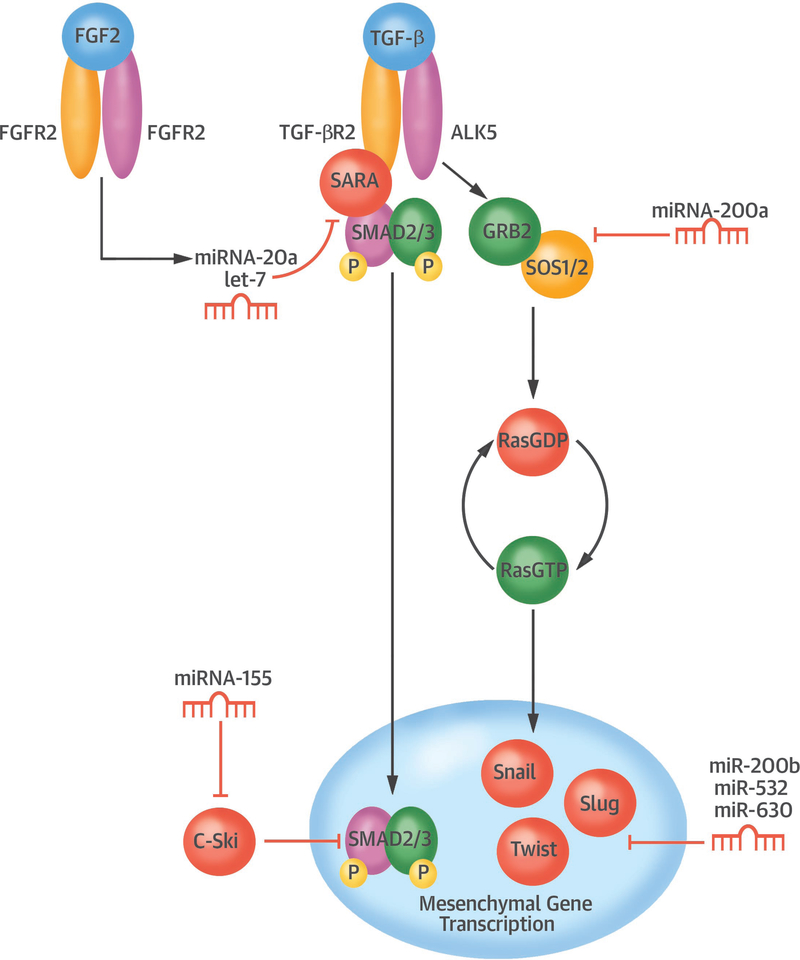
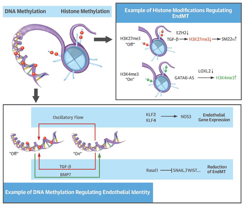
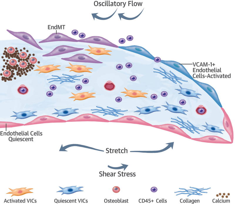
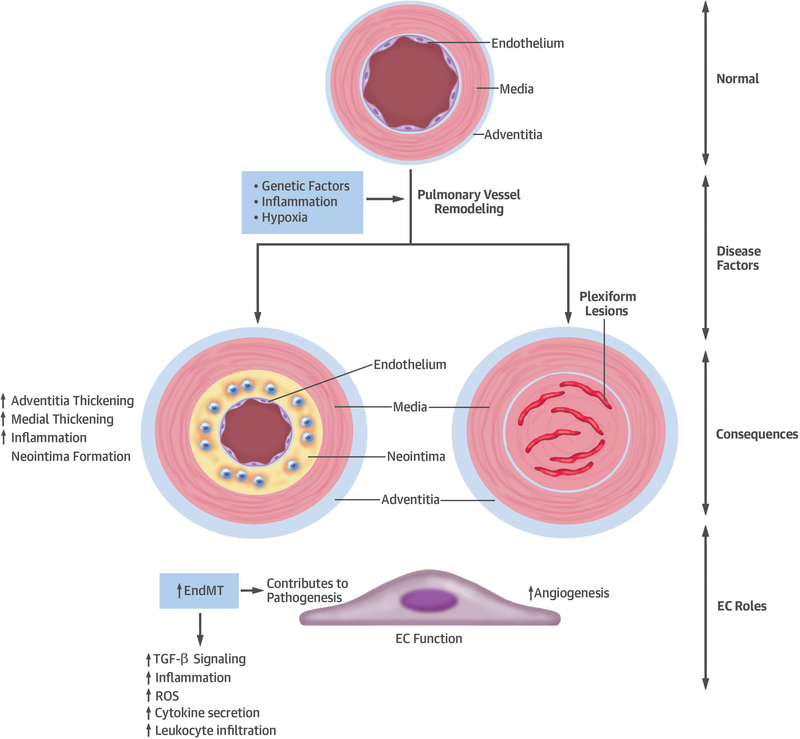
Endothelial to mesenchymal transition (EndMT) is a process whereby an endothelial cell undergoes a series of molecular events that lead to a change in phenotype toward a mesenchymal cell (e.g., myofibroblast, smooth muscle cell). EndMT plays a fundamental role during development, and mounting evidence indicates that EndMT is involved in adult cardiovascular diseases (CVDs), including atherosclerosis, pulmonary hypertension, valvular disease, and fibroelastosis. Therefore, the targeting of EndMT may hold therapeutic promise for treating CVD. However, the field faces a number of challenges, including the lack of a precise functional and molecular definition, a lack of understanding of the causative pathological role of EndMT in CVDs (versus being a "bystander-phenomenon"), and a lack of robust human data corroborating the extent and causality of EndMT in adult CVDs. Here, we review this emerging but exciting field, and propose a framework for its systematic advancement at the molecular and translational levels.

Keywords: EndMT; cardiovascular; endothelial to mesenchymal transition.


Conflict of interest statement

The authors declare no conflicts of interest.

Figures

Central Illustration. EndMT in CVD:
Central Illustration. EndMT in CVD:. Key Mechanisms and Clinical Translation Opportunities.
Summary of major concepts elucidated in this article. Adapted from (144) with permission.
Figure 1.
Figure 1.. EndMT during cardiac development.
(A) Schematic cross-section of the developing mouse heart at ~10.5 days showing major chambers, forming septa, and the outflow tract (OFT). (B) Schematic enlargement of one side of the AV canal (boxed region in A). During EndMT, ECs of the AV canal and OFT become mesenchymal and occupy the prominent ECM swellings separating myocardium and endocardium (endocardial cushions) in those regions. (C) Cellularized endocardial cushions are later remodeled into stratified valve leaflets. Here, a mitral valve leaflet is shown stratified into atrialis [A], spongiosa [S] and fibrosa [F] layers, and tethered to the ventricle by papillary muscles and chordae tendineae. (D) Complex signaling networks drive endocardial cushion formation, EndMT, expansion of cushion mesenchyme and cardiac remodeling, which are further modulated by biomechanical forces associated with heart contraction and blood flow. Key molecular factors (see text and Figure 2) and relevant human congenital and adult valvular diseases are indicated.
Figure 2.
Figure 2.. TGF-β signaling and EndMT.
In ECs, TGF-β classically signals via TGF-βR2 (a type II receptor component) and AKL1 or ALK5 (type I receptor complexes). Receptor complexes combine on the cell surface and are comprised of two type I and two type II components. TGF-βR2 phosphorylates (activates) type I components, which then propagate the signal intracellularly via activation (phosphorylation; pSMAD) of SMAD 1,2,3,5 or 8. Activated SMAD proteins form complexes that include the common mediator SMAD4, and which may be inhibited by SMAD6 or SMAD7. SMAD complexes shuttle to the nucleus, where they interact with co-activators, co-repressors and additional transcription factors, the latter including key EndMT gatekeepers SNAI1/2, ZEB1/2, KLF4, TCF3 and TWIST. These interactions culminate in chromatin rearrangements and transcription factor binding to endothelial, mesenchymal and other gene promoter regions that ultimately bring about EndMT.
Figure 3.
Figure 3.. Fatty acid oxidation regulates EndMT.
Key elements of the role of FAO in regulating EndMT.
Figure 4.
Figure 4.. The role of non-coding RNAs in regulating EndMT.
Key elements of what is known about how EndMT may be controlled by non-coding RNAs. Abbreviations not previously defined: FGFR, FGF receptor; SARA, SMAD anchor for receptor activation; SOS, Son of sevenless homolog.
Figure 5.
Figure 5.. Epigenetic mechanisms and control of EndMT.
Key elements of what is known of how epigenetic changes modulate EndMT.
Figure 6.
Figure 6.. EndMT in atherosclerosis and plaque erosion.
Confocal microscopy of thoracic aortic sections from tamoxifen-induced end.SclCreERT;R26RstopYfp;ApoE−/− mice after Western diet feeding. In this model of advanced atherosclerosis, ECs are permanently marked using a Cre-lox system such that ECs, and all EC-derived cells, permanently express yellow fluorescence protein (Yfp). Staining for Ve-Cadherin is in red, with staining for fibroblast activation protein (Fap), a fibroblast marker, in white. DAPI nuclear staining is in blue. Yfp+Fap+Ve-Cadherin+ cells (arrowheads) represent endothelial-derived cells expressing endothelial and fibroblast proteins. Yfp+Fap+Ve-Cadherin- cells (arrows) represent endothelial-derived cells that express Fap, but which have lost Ve-Cadherin expression. L=lumen; scale bars, 100 μm. Reproduced from (8) with permission.
Figure 7.
Figure 7.. EndMT in heart valve disease.
In normal valves, valvular interstitial cells (VICs) possess a quiescent phenotype. VICs become activated in disease. EndMT in heart valves could be initiated by mechanical stress and inflammation (CD45-positive cells). Activated VCAM1-positive endothelium undergoes EndMT, which generates more interstitial cells. Some of these cells may undergo osteogenic transformation and activation resulting in various diseases.
Figure 8.
Figure 8.. EndMT in PAH.
Key features of PAH and the role of EndMT.

Similar articles

Cited by

References

    1. Hamburger V Introduction: Johannes Holtfreter, pioneer in experimental embryology. Dev Dyn 1996;205:214–6. - PubMed
    1. Trelstad RL, Hay ED, Revel JD. Cell contact during early morphogenesis in the chick embryo. Dev Biol 1967;16:78–106. - PubMed
    1. Kovacic JC, Mercader N, Torres M, Boehm M, Fuster V. Epithelial-to-mesenchymal and endothelial-to-mesenchymal transition: from cardiovascular development to disease. Circulation 2012;125:1795–808. - PMC - PubMed
    1. Grande MT, Sanchez-Laorden B, Lopez-Blau C et al. Snail1-induced partial epithelial-to-mesenchymal transition drives renal fibrosis in mice and can be targeted to reverse established disease. Nat Med 2015;21:989–97. - PubMed
    1. Santamaria PG, Moreno-Bueno G, Portillo F, Cano A. EMT: Present and future in clinical oncology. Molecular oncology 2017;11:718–738. - PMC - PubMed

Publication types