

Ionotropic Receptors Specify the Morphogenesis of Phasic Sensors Controlling Rapid Thermal Preference in Drosophila
- PMID: 30654923
- PMCID: PMC6709853
- DOI: 10.1016/j.neuron.2018.12.022
Ionotropic Receptors Specify the Morphogenesis of Phasic Sensors Controlling Rapid Thermal Preference in Drosophila
Abstract
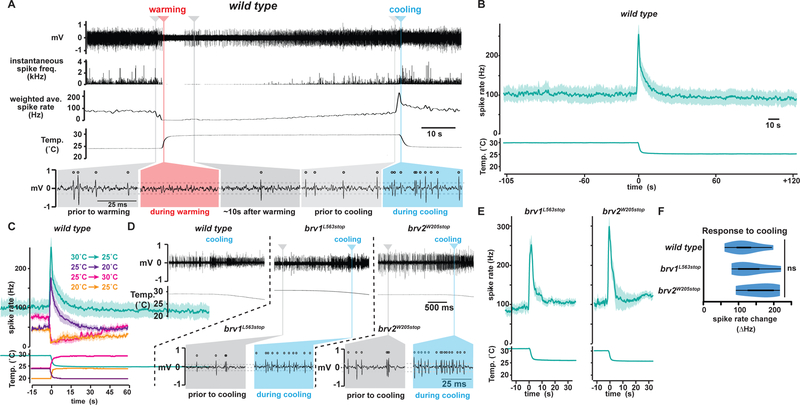
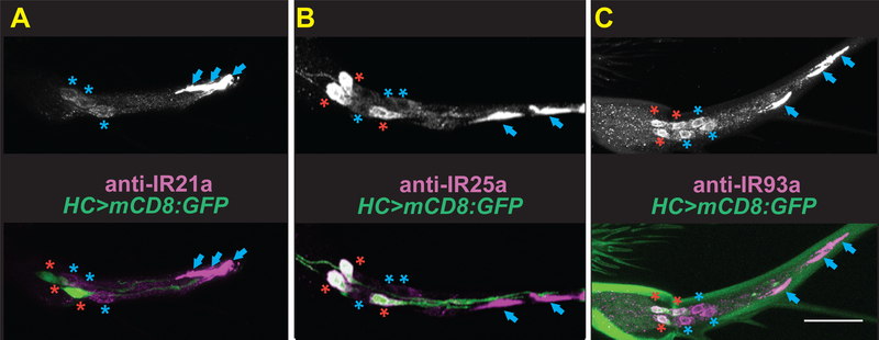
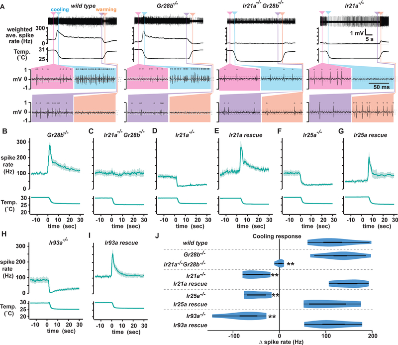
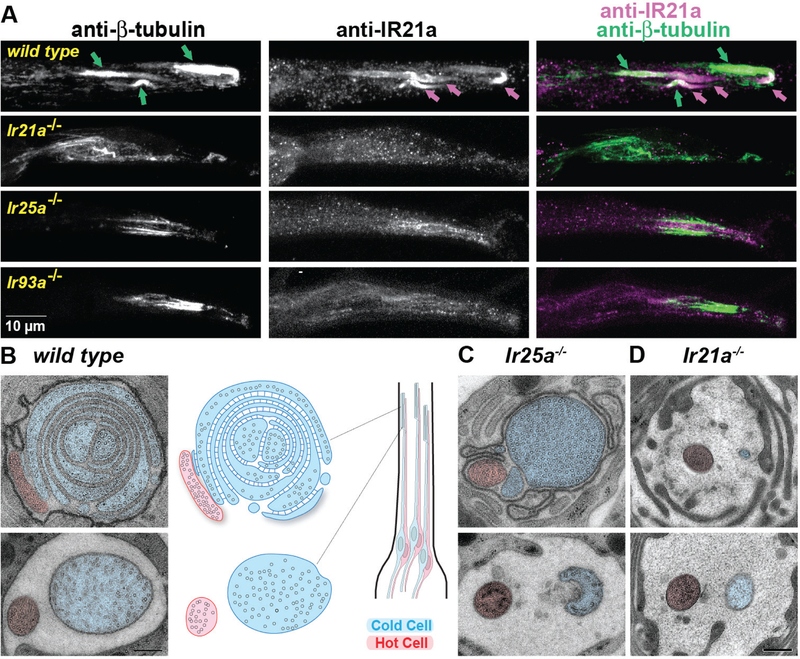
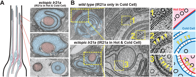
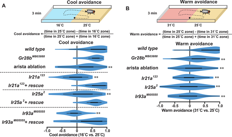
Thermosensation is critical for avoiding thermal extremes and regulating body temperature. While thermosensors activated by noxious temperatures respond to hot or cold, many innocuous thermosensors exhibit robust baseline activity and lack discrete temperature thresholds, suggesting they are not simply warm and cool detectors. Here, we investigate how the aristal Cold Cells encode innocuous temperatures in Drosophila. We find they are not cold sensors but cooling-activated and warming-inhibited phasic thermosensors that operate similarly at warm and cool temperatures; we propose renaming them "Cooling Cells." Unexpectedly, Cooling Cell thermosensing does not require the previously reported Brivido Transient Receptor Potential (TRP) channels. Instead, three Ionotropic Receptors (IRs), IR21a, IR25a, and IR93a, specify both the unique structure of Cooling Cell cilia endings and their thermosensitivity. Behaviorally, Cooling Cells promote both warm and cool avoidance. These findings reveal a morphogenetic role for IRs and demonstrate the central role of phasic thermosensing in innocuous thermosensation. VIDEO ABSTRACT.
Keywords: Ir21a; Ir25a; Ir93a; iGluR; ionotropic receptor; morphogenesis; sensory neuron; temperature; thermoreceptor; thermosensation.
Copyright © 2018 Elsevier Inc. All rights reserved.
Conflict of interest statement
Figures
Comment in
-
A Fly's Cool Way to Escape the Heat.Neuron. 2019 Feb 20;101(4):550-552. doi: 10.1016/j.neuron.2019.01.054. Neuron. 2019. PMID: 30790534
Similar articles
-
The Ionotropic Receptors IR21a and IR25a mediate cool sensing in Drosophila.Elife. 2016 Apr 29;5:e13254. doi: 10.7554/eLife.13254. Elife. 2016. PMID: 27126188 Free PMC article.
-
Monoacylglycerol acyltransferase maintains ionotropic receptor expression for cool temperature sensing and avoidance in Drosophila.Commun Biol. 2025 May 29;8(1):765. doi: 10.1038/s42003-025-08154-0. Commun Biol. 2025. PMID: 40442407 Free PMC article.
-
Distinct combinations of variant ionotropic glutamate receptors mediate thermosensation and hygrosensation in Drosophila.Elife. 2016 Sep 22;5:e17879. doi: 10.7554/eLife.17879. Elife. 2016. PMID: 27656904 Free PMC article.
-
Ionotropic receptors (IRs): chemosensory ionotropic glutamate receptors in Drosophila and beyond.Insect Biochem Mol Biol. 2013 Sep;43(9):888-97. doi: 10.1016/j.ibmb.2013.02.007. Epub 2013 Mar 1. Insect Biochem Mol Biol. 2013. PMID: 23459169 Review.
-
Trp ion channels and temperature sensation.Annu Rev Neurosci. 2006;29:135-61. doi: 10.1146/annurev.neuro.29.051605.112958. Annu Rev Neurosci. 2006. PMID: 16776582 Review.
Cited by
-
Ionotropic Receptor-dependent cool cells control the transition of temperature preference in Drosophila larvae.PLoS Genet. 2021 Apr 7;17(4):e1009499. doi: 10.1371/journal.pgen.1009499. eCollection 2021 Apr. PLoS Genet. 2021. PMID: 33826603 Free PMC article.
-
An integrated anatomical, functional and evolutionary view of the Drosophila olfactory system.EMBO Rep. 2025 Jun;26(12):3204-3225. doi: 10.1038/s44319-025-00476-8. Epub 2025 May 19. EMBO Rep. 2025. PMID: 40389758 Free PMC article.
-
Cold-Sensing TRP Channels and Temperature Preference Modulate Ovarian Development in the Model Organism Drosophila melanogaster.Int J Mol Sci. 2025 Jun 12;26(12):5638. doi: 10.3390/ijms26125638. Int J Mol Sci. 2025. PMID: 40565102 Free PMC article.
-
Temperature synchronization of the Drosophila circadian clock protein PERIOD is controlled by the TRPA channel PYREXIA.Commun Biol. 2019 Jul 1;2:246. doi: 10.1038/s42003-019-0497-0. eCollection 2019. Commun Biol. 2019. PMID: 31286063 Free PMC article.
-
Functional labeling of individualized postsynaptic neurons using optogenetics and trans-Tango in Drosophila (FLIPSOT).PLoS Genet. 2024 Mar 14;20(3):e1011190. doi: 10.1371/journal.pgen.1011190. eCollection 2024 Mar. PLoS Genet. 2024. PMID: 38483970 Free PMC article.
References
-
- Altner H, and Loftus R (1985). Ultrastructure and function of insect thermo- and hygroreceptors. Ann Rev Entomol 30, 273–295.
-
- Biron D, Wasserman S, Thomas JH, Samuel AD, and Sengupta P (2008). An olfactory neuron responds stochastically to temperature and modulates Caenorhabditis elegans thermotactic behavior. Proceedings of the National Academy of Sciences of the United States of America 105, 11002–11007. doi: 10.1073/pnas.0805004105 - DOI - PMC - PubMed
Publication types
MeSH terms
Substances
Grants and funding
LinkOut - more resources
Full Text Sources
Molecular Biology Databases
