. 2019 Mar 15;294(11):3947-3956.
doi: 10.1074/jbc.RA118.006391. Epub 2019 Jan 17.

The p12 subunit of human polymerase δ uses an atypical PIP box for molecular recognition of proliferating cell nuclear antigen (PCNA)

Affiliations

The p12 subunit of human polymerase δ uses an atypical PIP box for molecular recognition of proliferating cell nuclear antigen (PCNA)

Amaia Gonzalez-Magaña et al. J Biol Chem. .

Abstract

Human DNA polymerase δ is essential for DNA replication and acts in conjunction with the processivity factor proliferating cell nuclear antigen (PCNA). In addition to its catalytic subunit (p125), pol δ comprises three regulatory subunits (p50, p68, and p12). PCNA interacts with all of these subunits, but only the interaction with p68 has been structurally characterized. Here, we report solution NMR-, isothermal calorimetry-, and X-ray crystallography-based analyses of the p12-PCNA interaction, which takes part in the modulation of the rate and fidelity of DNA synthesis by pol δ. We show that p12 binds with micromolar affinity to the classical PIP-binding pocket of PCNA via a highly atypical PIP box located at the p12 N terminus. Unlike the canonical PIP box of p68, the PIP box of p12 lacks the conserved glutamine; binds through a 2-fork plug made of an isoleucine and a tyrosine residue at +3 and +8 positions, respectively; and is stabilized by an aspartate at +6 position, which creates a network of intramolecular hydrogen bonds. These findings add to growing evidence that PCNA can bind a diverse range of protein sequences that may be broadly grouped as PIP-like motifs as has been previously suggested.

Keywords: DNA polymerase; DNA replication; PCNA interacting sequence; PIP-box; processivity; proliferating cell nuclear antigen (PCNA); protein structure; protein-protein interaction.


Conflict of interest statement

The authors declare that they have no conflicts of interest with the contents of this article

Figures

Figure 1.
Figure 1.
Amino acid sequence of p12 together with disorder and secondary structure predictions. Residues encompassing the peptide used for crystallization and biophysical characterization (p121–19) with PCNA are indicated in bold pink characters, and those belonging to the divergent PCNA interacting motif are boxed. The consensus residues in the PIP-like motif are indicated by arrows. The solid line shows the disorder prediction by the PrDOS server (51), and the red line shows the threshold of 0.5. Secondary structure elements (helices) predicted by JPred4 (52) are indicated above the sequence.
Figure 2.
Figure 2.
NMR analysis of the p12 peptide interaction with PCNA. A, superposition of 1H-15N TROSY spectra of 51 μm PCNA in the absence (black) and presence (red) of a 10-fold molar excess of p121–19 peptide. Spectra were acquired at 35 °C in PBS, pH 7.0, 1 mm DTT. B, region of the NMR spectra of PCNA in the presence of increasing amounts of p12 peptide (from black to red) showing the titration of Leu235 signal. C, CSPs of PCNA backbone amide 1H and 15N NMR resonances induced by p121–19. The dashed line indicates the average plus one standard deviation. The green bars indicate the positions of residues that disappear upon peptide addition. D, chemical shift perturbation of the amide signal of residues with CSP larger than the average plus one standard deviation at different p12:PCNA ratios. The symbols correspond to the experimental data, and the lines correspond to the best fits to a model of one set of identical binding sites.
Figure 3.
Figure 3.
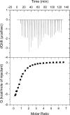
Isothermal calorimetric titration of PCNA with p12 peptide. The upper panel represents the heat effect associated with the variable volume peptide injection, and the lower panel represents the ligand concentration dependence of the heat released upon binding, after normalization and correction for the heat of dilution. The molar ratio is that of p12:PCNA protomer. The symbols correspond to the experimental data, and the continuous line corresponds to the best fit to a model of one set of identical binding sites.
Figure 4.
Figure 4.
Crystal structure of the p12–PCNA complex and comparison with p68–PCNA structure. A, overall structure of trimeric human PCNA (green) bound to the peptide derived from p12 (p121–19, magenta). Only p12 residues 3–15 are seen in the electron density map. B, p12–PCNA–binding site highlighting p12 intramolecular interactions. PCNA surface is shown in pale green, the p12 peptide is in magenta with stick representation, and interactions are shown as yellow dotted lines. Peptide residues involved in the interactions are labeled. 2FoFc map around the p12 peptide is shown in blue contoured at 1 σ. C, p12–PCNA–binding site highlighting p12 intermolecular interactions. PCNA is shown in green with ribbon representation, p12 is in magenta with stick representation, and interactions are shown as yellow dotted lines. PCNA and p12 interacting residues are labeled. Residues of PCNA involved in hydrophobic interactions are boxed. D, p68–PCNA–binding site (PDB code 1U76 (20)). The PCNA surface is shown in dark gray, and p68 (residues 453–465) is in turquoise stick representation. The p68 PIP-box consensus residues are labeled. C-term, C-terminus.
Figure 5.
Figure 5.
NMR analysis of the RecQ5 peptide interaction with PCNA. A, superposition of 1H-15N TROSY spectra of 50 μm PCNA in the absence (black) and presence (red) of a 37-fold molar excess of RecQ5 peptide. The spectra were acquired at 35 °C in PBS, pH 7.0, 1 mm DTT. B, CSPs of PCNA backbone amide 1H and 15N NMR resonances induced by RecQ5952–979. The dotted line indicates the average plus one standard deviation. The green bars indicate the positions of residues that disappear upon peptide addition. C, chemical shift perturbation of the amide signal of PCNA residues with CSPs larger than the average plus one standard deviation at different RecQ5:PCNA ratios. The symbols correspond to the experimental data, and the lines correspond to the best fit to a model of one set of identical binding sites.
Figure 6.
Figure 6.
NMR chemical shift mapping of the RecQ5 (A) or p12 (B) interaction site on PCNA. Front and back views of one of the PCNA protomers are shown in white surface representation, whereas p12 peptide in the crystallographic position is shown as blue sticks. PCNA residues whose amide signals significantly shift, disappear, or remain unassigned in the titration with the RecQ5 or p12 peptides are painted in red, orange, or gray, respectively.
Figure 7.
Figure 7.
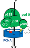
Possible organization of the human pol δ–PCNA complex on primer/template DNA. The N-terminal (N-t) and C-terminal (C-t) PIP motifs of p12 and p68 subunits, connected to the folded domains by disordered regions (shown as dashed lines), are indicated with red or yellow boxes, respectively, and binding two distinct PCNA subunits; CTD, p125 C-terminal Domain.
Figure 8.
Figure 8.
A, comparison of p21 and p12 PIP degrons interacting with PCNA. B, superposition of structures of canonical (left) and noncanonical (right) PCNA-interacting motifs bound to PCNA. A, p21–PCNA (PDB code 1AXC) (22) and p12–PCNA (PDB code 6HVO; current study) structures are aligned. p21 and p12 peptides are shown as yellow and magenta sticks, respectively. PCNA is shown as a green surface. The residues making up the acidic patch in the IDCL are colored red. B, left panel, the PCNA protomers are represented by ribbons, and the peptides are represented by their Cα traces. The color code is as follows: p21, yellow (PDB code 1AXC) (22); p15PAF, red (PDB code 4D2G) (44); FEN-1, blue (PDB code 1U7B) (20); p68, green (PDB code 1U76) (20); ZRANB3-PIP, purple (PDB code 5MLO) (42); DVC1, gray (PDB code 5IY4) (53). B, right panel, the color code is as follows: PARG, gray (PDB code 5MAV) (38); ZRANB3-APIM, brown (PDB code 5MLW) (41); pol η, blue (PDB code 2ZVK) (37); pol ı, purple (PDB code 2ZVM) (37); pol κ, yellow (PDB code 2ZVL) (37); RNH2B, green (PDB code 3P87) (54); TRAIP, red (PDB code 4ZTD) (29); and p12, orange (PDB code 6HVO).

References

    1. Hubscher U., Maga G., and Spadari S. (2002) Eukaryotic DNA polymerases. Annu. Rev. Biochem. 71, 133–163 10.1146/annurev.biochem.71.090501.150041 - DOI - PubMed
    1. Lee M., Wang X., Zhang S., Zhang Z., and Lee E. Y. C. (2017) Regulation and modulation of human DNA polymerase δ activity and function. Genes (Basel) 8, E190 10.3390/genes8070190 - DOI - PMC - PubMed
    1. Zhang S., Zhou Y., Trusa S., Meng X., Lee E. Y., and Lee M. Y. (2007) A novel DNA damage response: rapid degradation of the p12 subunit of DNA polymerase δ. J. Biol. Chem. 282, 15330–15340 10.1074/jbc.M610356200 - DOI - PubMed
    1. Meng X., Zhou Y., Zhang S., Lee E. Y., Frick D. N., and Lee M. Y. (2009) DNA damage alters DNA polymerase δ to a form that exhibits increased discrimination against modified template bases and mismatched primers. Nucleic Acids Res. 37, 647–657 - PMC - PubMed
    1. Meng X., Zhou Y., Lee E. Y., Lee M. Y., and Frick D. N. (2010) The p12 subunit of human polymerase δ modulates the rate and fidelity of DNA synthesis. Biochemistry 49, 3545–3554 10.1021/bi100042b - DOI - PMC - PubMed

Publication types

MeSH terms

Substances