. 2019 Apr 15;25(8):2560-2574.
doi: 10.1158/1078-0432.CCR-18-0432. Epub 2019 Jan 17.

CAR T Cells Targeting B7-H3, a Pan-Cancer Antigen, Demonstrate Potent Preclinical Activity Against Pediatric Solid Tumors and Brain Tumors

Affiliations

CAR T Cells Targeting B7-H3, a Pan-Cancer Antigen, Demonstrate Potent Preclinical Activity Against Pediatric Solid Tumors and Brain Tumors

Robbie G Majzner et al. Clin Cancer Res. .

Abstract

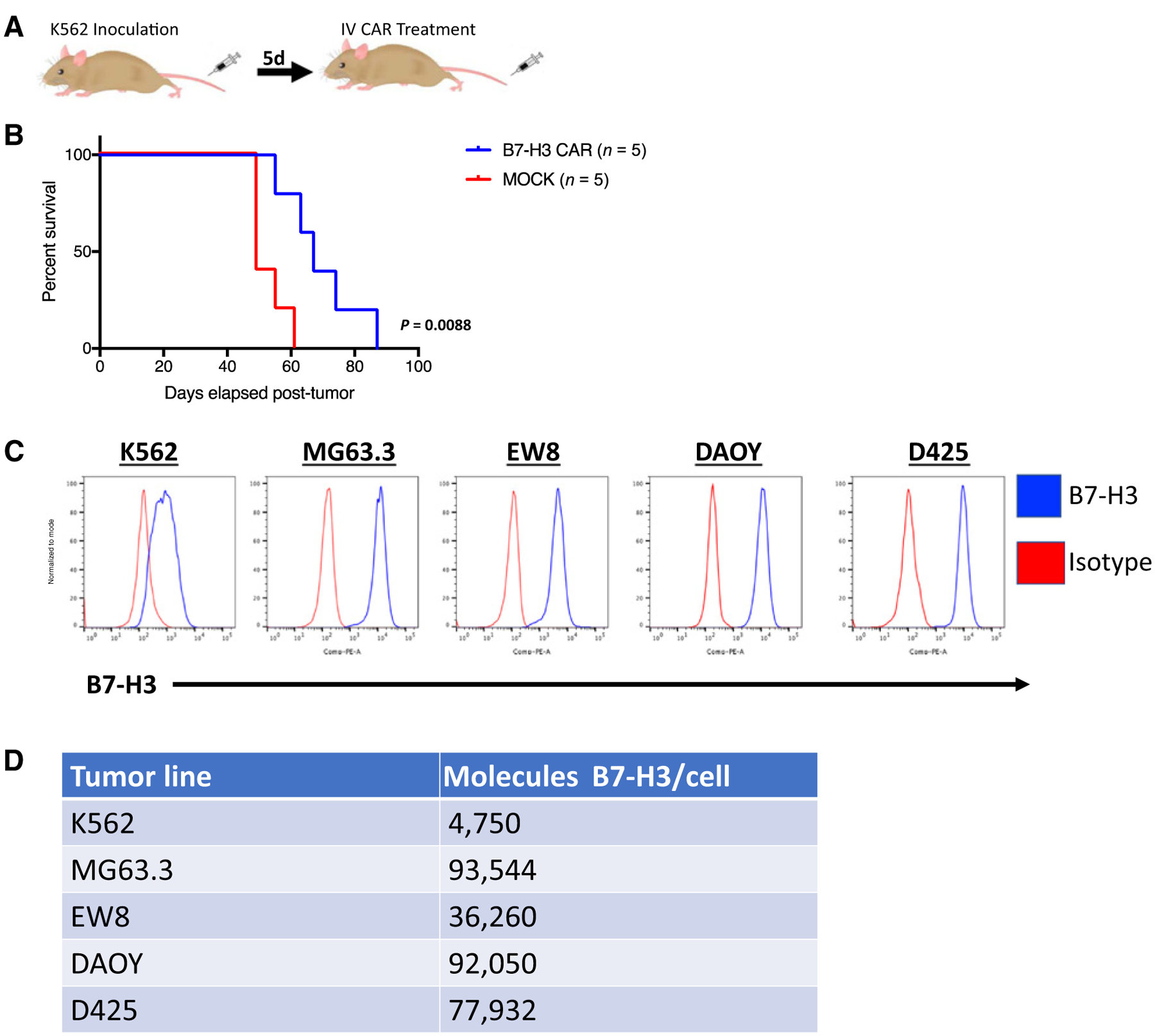
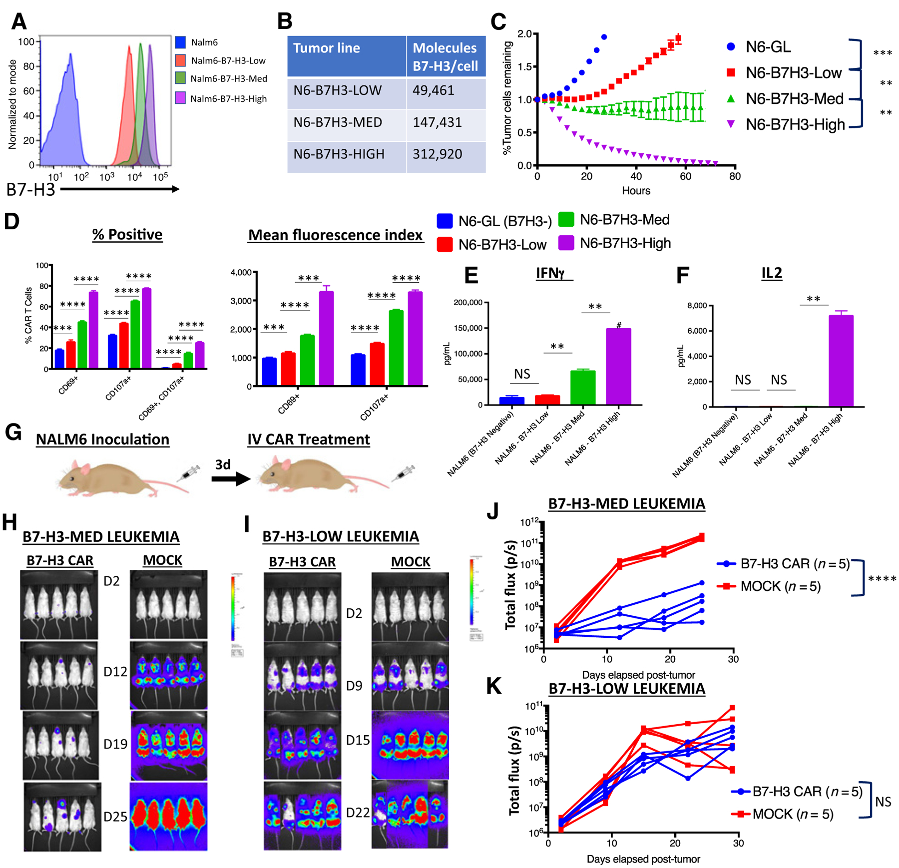
Purpose: Patients with relapsed pediatric solid tumors and CNS malignancies have few therapeutic options and frequently die of their disease. Chimeric antigen receptor (CAR) T cells have shown tremendous success in treating relapsed pediatric acute lymphoblastic leukemia, but this has not yet translated to treating solid tumors. This is partially due to a paucity of differentially expressed cell surface molecules on solid tumors that can be safely targeted. Here, we present B7-H3 (CD276) as a putative target for CAR T-cell therapy of pediatric solid tumors, including those arising in the central nervous system.

Experimental design: We developed a novel B7-H3 CAR whose binder is derived from a mAb that has been shown to preferentially bind tumor tissues and has been safely used in humans in early-phase clinical trials. We tested B7-H3 CAR T cells in a variety of pediatric cancer models.

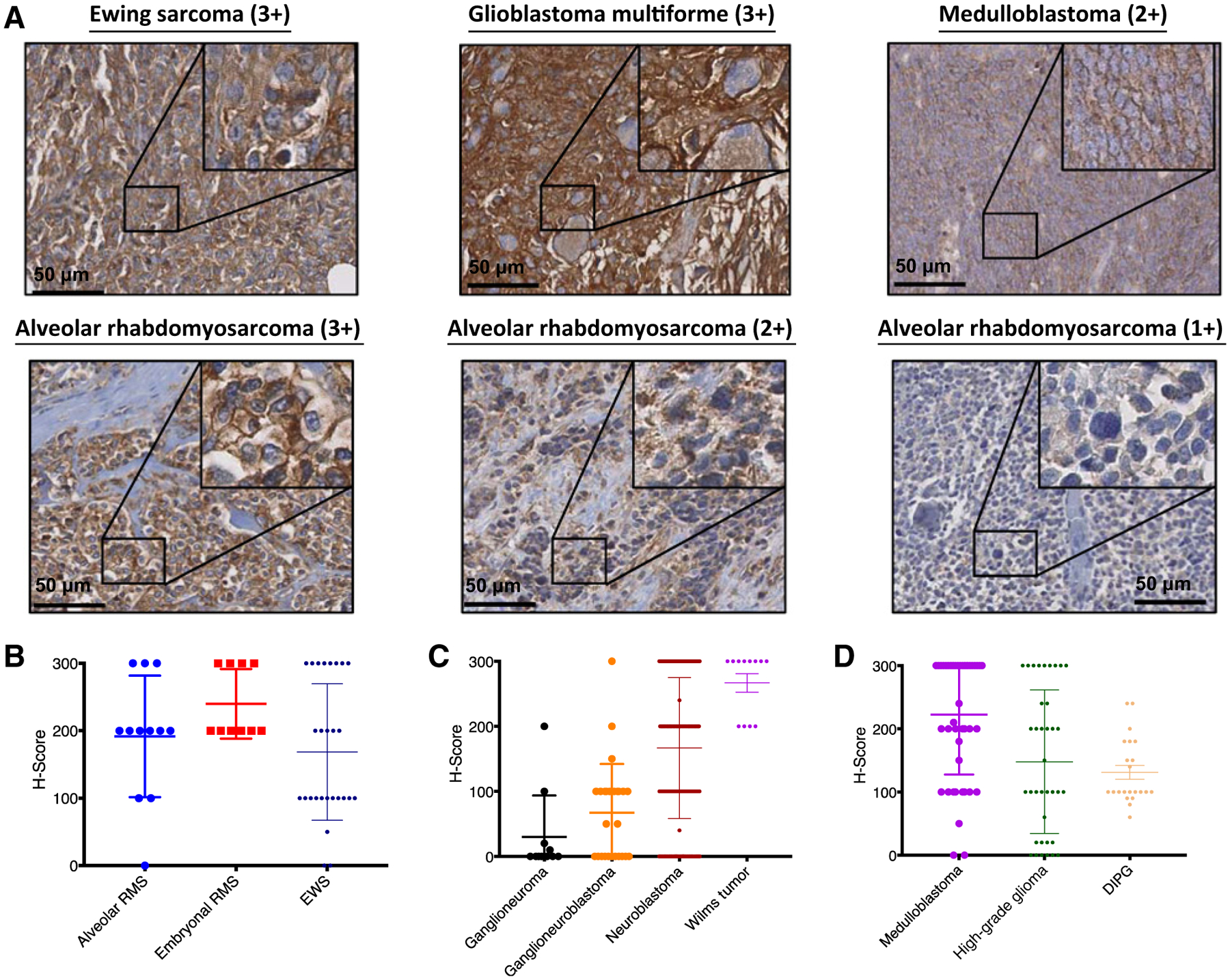
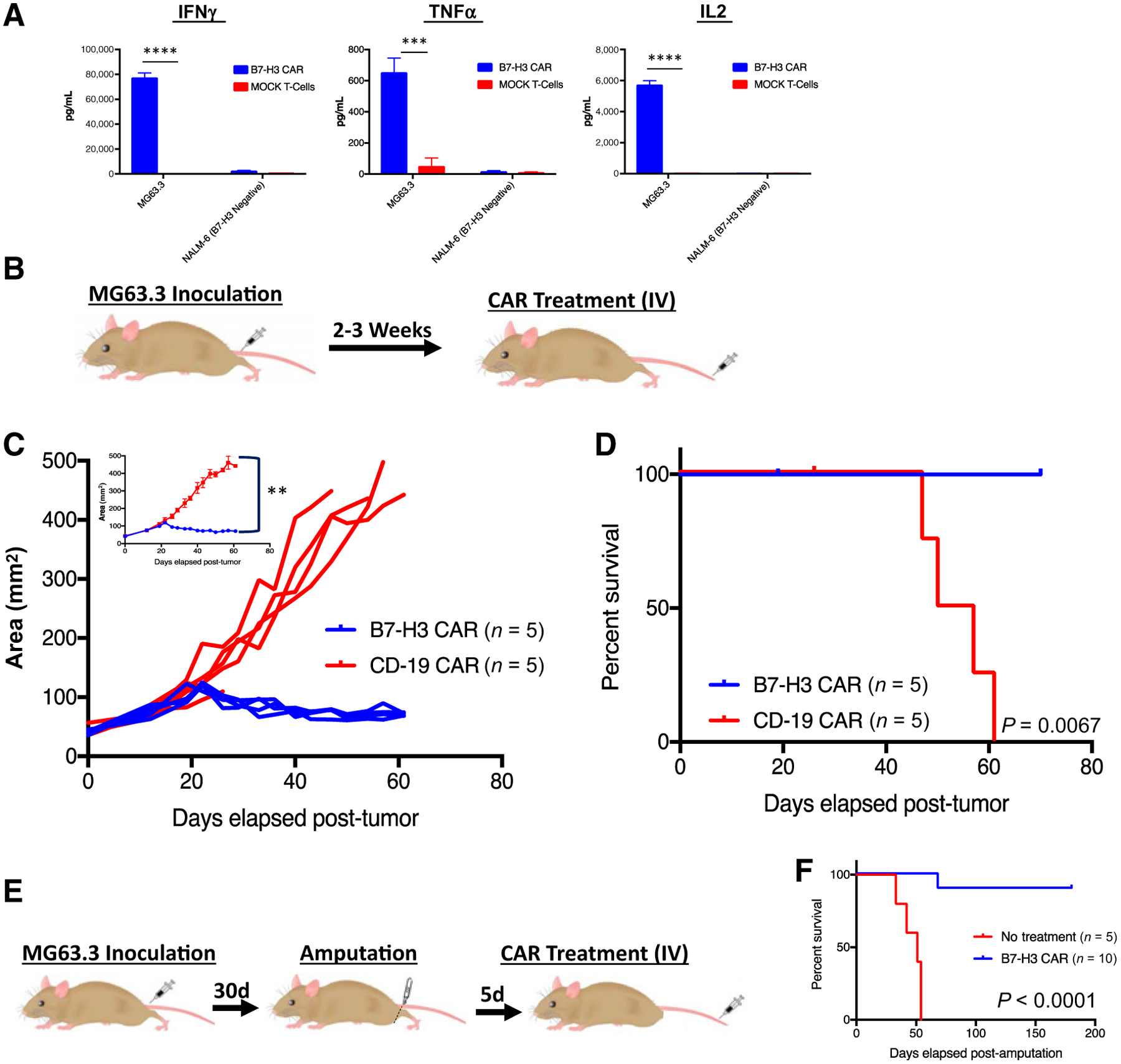
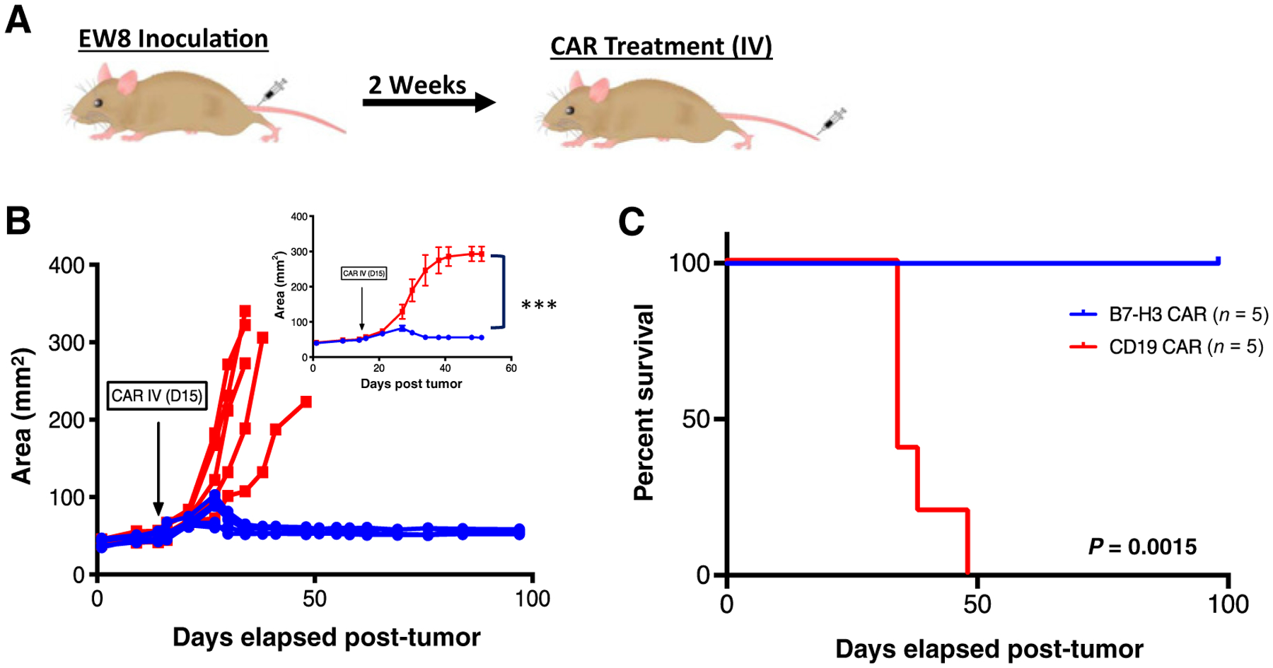
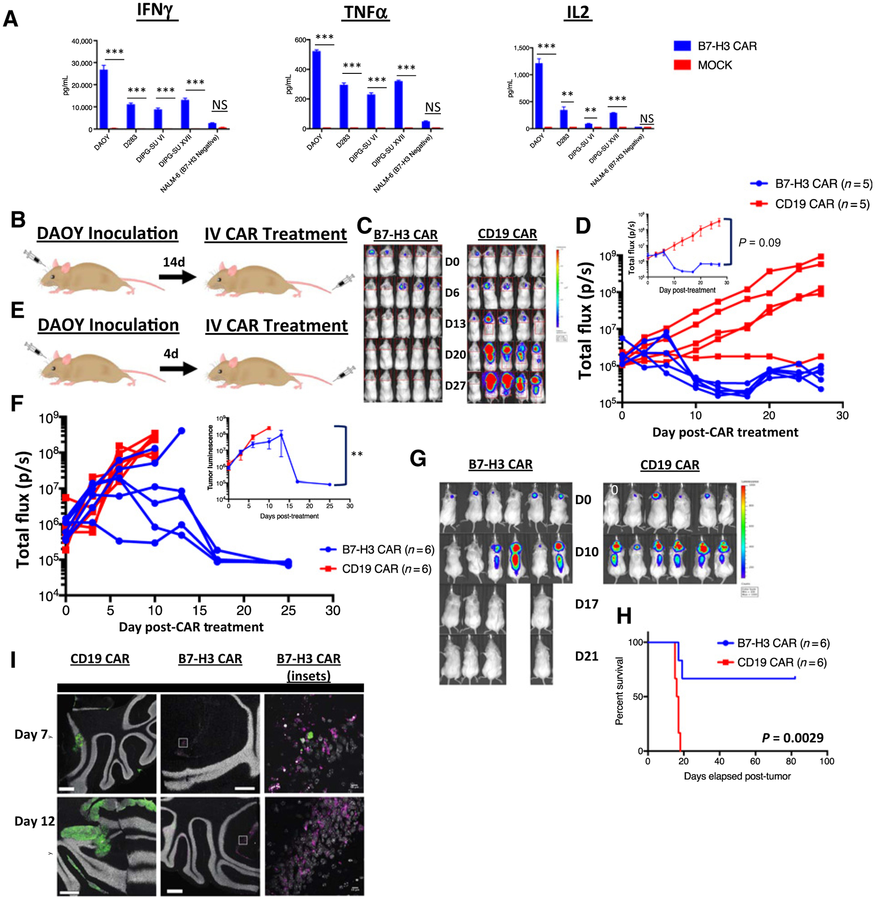
Results: B7-H3 CAR T cells mediate significant antitumor activity in vivo, causing regression of established solid tumors in xenograft models including osteosarcoma, medulloblastoma, and Ewing sarcoma. We demonstrate that B7-H3 CAR T-cell efficacy is largely dependent upon high surface target antigen density on tumor tissues and that activity is greatly diminished against target cells that express low levels of antigen, thus providing a possible therapeutic window despite low-level normal tissue expression of B7-H3.

Conclusions: B7-H3 CAR T cells could represent an exciting therapeutic option for patients with certain lethal relapsed or refractory pediatric malignancies, and should be tested in carefully designed clinical trials.


Figures

Figure 1.
Figure 1.
B7-H3 is highly expressed on pediatric solid tumors. A, Pediatric tumor microarrays were stained by IHC for the expression of B7-H3. Representative images of Ewing sarcoma (3+), glioblastoma multiforme (3+), medulloblastoma (2+), and alveolar rhabdomyosarcoma (3+, 2+, and 1+) samples are shown. H-scores were generated by multiplying the % cells positive × intensity seen for each core. H-scores are shown for pediatric sarcomas (B), neuroblastoma and Wilms tumor (C), and pediatric CNS tumors (D). RMS, rhabdomyosarcoma; EWS, Ewing sarcoma; DIPG, diffuse intrinsic pontine glioma.
Figure 2.
Figure 2.
Systemically administered B7-H3 CAR T cells induce regression of osteosarcoma xenografts. A, B7-H3 CAR T cells produce IFNγ, TNFα, and IL2 following 24-hour in vitro coculture with MG63.3 osteosarcoma. Representative results of four experiments with 3 different PBMC donors are shown. B, Mouse model of orthotopic osteosarcoma: 1e6 MG63.3 tumor cells were injected into the periosteum of the tibia in NSG mice. Two to three weeks later, following onset of measurable tumors, 1e7 B7-H3 CAR+ T cells or irrelevant control CD19 CAR T cells were intravenously administered. C, Tumor growth was measured biweekly by digital caliper and tumor area was calculated. Values for individual mice, as well as mean values per treatment group (inset) are shown. D, Survival curves of mice treated as in B. Representative results of three experiments with 3 different PBMC donors are shown. E, Metastatic model of osteosarcoma: MG63.3-derived tumors were allowed to grow and metastasize before the mouse underwent amputation followed by administration of intravenous 1e7 B7-H3 CAR+ T cells. F, Survival curves of mice treated as in E. Representative results of four experiments with 3 different PBMC donors are shown. Error bars, SEM. P values were calculated as described in Materials and Methods.
Figure 3.
Figure 3.
Systemically administered B7-H3 CAR T cells induce regression of Ewing sarcoma xenografts. A, Mouse model of orthotopic Ewing sarcoma: 2e7 EW8 tumor cells were injected into the periosteum of the tibia in NSG mice. Two weeks later, 1e7 B7-H3 CAR+ T cells or irrelevant control CD19 CAR T cells were intravenously administered. B, Tumor growth was measured twice weekly by digital caliper and tumor area was calculated. Values for individual mice, as well as mean values per treatment group (inset) are shown. C, Survival curves of mice treated as in A. Representative results of two experiments with 2 different PBMC donors are shown. Error bars, SEM. P values were calculated as described in Materials and Methods.
Figure 4.
Figure 4.
Systemically administered B7-H3 CAR T cells can clear medulloblastoma xenografts. A, B7-H3 CAR T cells were cocultured in vitro with medulloblastoma cell lines and patient-derived DIPG cell cultures and, 24 hours later, IFNγ, TNFα, and IL2 were measured in the supernatant. Representative results of three experiments with 3 different PBMC donors are shown. B, Orthotopic xenograft model of medulloblastoma: NSG mice were autochthonously injected with luciferase expressing DAOY medulloblastoma tumor cells. Following evidence of tumor engraftment by IVIS imaging, animals received 1e7 B7-H3 CAR+ T cells or CD19 CAR T cells intravenously. C, In vivo imaging of DAOY tumors treated with B7-H3 or CD19 CAR T cells. D, Tumor progression was measured by bioluminescence photometry and flux values (photons per second) were calculated using Living Image software. Values for individual mice, as well as mean values per treatment group (inset) are shown. Representative results of three experiments with three different PBMC donors are shown. E, Orthotopic xenograft model of c-myc–amplified medulloblastoma: D425 tumor cells expressing luciferase were autochthonously injected into NSG mice. Mice were treated with 1e7 B7-H3 CAR+ T cells or CD19 CAR T cells after 3–4 days, at which point tumor was detectable by IVIS imaging. F, Tumor progression was measured by bioluminescence photometry and flux values (photons per second) were calculated using Living Image software. Values for individual mice, as well as average values of living mice per treatment group (inset) are shown. G, In vivo imaging of D425 tumors treated with B7-H3 or CD19 CAR T cells. H, Survival curves of mice shown in G. Representative results of three experiments with three different T cell donors are shown. I, Confocal images of brains from D425-GFP+ tumor bearing mice treated with B7-H3 CAR-mCherry or CD19 CAR-mCherry T cells, harvested at two different time points after T-cell infusion. Representative image of two mice at two time points in one experiment. Error bars, SEM. P values were calculated as described in Materials and Methods.
Figure 5.
Figure 5.
B7-H3 CAR T cells have limited activity against B7-H3 low expressing K562 xenografts. A, Mouse model of K562 leukemia: NSG mice were inoculated with K562, a myeloid leukemia that expresses low levels of B7-H3, and then treated with 1e7 B7-H3 CAR+ T cells or mock transduced control T cells 5 days later. B, Survival curves of mice treated as in A. Representative results of five experiments with three different PBMC donors are shown. C, Flow cytometric analysis of B7-H3 expression on the surface of K562 (leukemia), MG63.3 (osteosarcoma), EW8 (Ewing sarcoma), DAOY, and D425 (medulloblastoma) human cell lines. D, Number of B7-H3 surface molecules expressed by human tumor cell lines as estimated by Quantibrite kit.
Figure 6.
Figure 6.
B7-H3 CAR T cells require adequate antigen expression for in vitro and in vivo activity. A, Flow cytometry analysis of B7-H3 expression on single-cell clones derived from Nalm6 expressing different amounts of lentivirally expressed B7-H3. B, Number of B7-H3 surface molecules expressed by Nalm6-B7-H3 cell lines as estimated by Quantibrite kit. C, GFP+ Nalm6-B7H3 clones were cocultured with B7-H3 CAR T cells and tumor cell killing was measured in an Incucyte assay over 72 hours. Representative data of three experiments with three different PBMC donors is shown. D, Percentage of CAR T cells positive (left) and mean fluorescence index (right) for T-cell activation and degranulation markers CD69 and CD107a, as measured by flow cytometry 6 hours after coculture of B7-H3 CAR T cells with tumor cells expressing increasing amounts of B7-H3. Representative results of three experiments with three different PBMC donors are shown. E and F, Cytokine production by CAR T cells cocultured with tumor cells expressing increasing amounts of B7-H3. G, Mouse model for Nalm6-B7H3: 1e6 NALM6 cells expressing either low or medium amounts of B7-H3 were engrafted into mice by tail vein injection. Three days later, mice were injected with 1e7 B7-H3 CAR+ T cells or untransduced control T cells (MOCK). In vivo imaging of mice bearing (H) Nalm6-B7-H3-Medium leukemia or (I) Nalm6-B7-H3-Low leukemia. J and K, Tumor progression was measured by bioluminescence photometry and flux values (photons per second) were calculated using Living Image software. Values for individual mice are shown. Representative results of four (Nalm6-B7-H3-Med) and two (Nalm6-B7-H3-Low) experiments with two different PBMC donors are shown. N6, NALM6; GL, GFP-Luciferase.

Similar articles

Cited by

References

    1. Smith MA, Altekruse SF, Adamson PC, Reaman GH, Seibel NL. Declining childhood and adolescent cancer mortality. Cancer 2014;120:2497–506. - PMC - PubMed
    1. Perkins SM, Shinohara ET, DeWees T, Frangoul H. Outcome for children with metastatic solid tumors over the last four decades. PLoS One 2014;9: e100396. - PMC - PubMed
    1. Packer RJ. Childhood brain tumors: accomplishments and ongoing challenges. J Child Neurol 2008;23:1122–7. - PMC - PubMed
    1. Rodriguez-Galindo C, Billups CA, Kun LE, Rao BN, Pratt CB, Merchant TE, et al.Survival after recurrence of Ewing tumors: the St Jude Children’s Research Hospital experience, 1979–1999. Cancer 2002;94:561–9. - PubMed
    1. Tomlinson FH, Scheithauer BW, Meyer FB, Smithson WA, Shaw EG, Miller GM, et al.Medulloblastoma: I. Clinical, diagnostic, and therapeutic overview. J Child Neurol 1992;7:142–55. - PubMed

Publication types

MeSH terms