Precision Cardio-Oncology
- PMID: 30655328
- PMCID: PMC6448460
- DOI: 10.2967/jnumed.118.220137
Precision Cardio-Oncology
Abstract
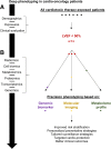
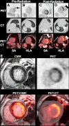
Modern oncologic therapies and care have resulted in a growing population of cancer survivors with comorbid, chronic health conditions. As an example, many survivors have an increased risk of cardiovascular complications secondary to cardiotoxic systemic and radiation therapies. In response, the field of cardio-oncology has emerged as an integral component of oncologic patient care, committed to the early diagnosis and treatment of adverse cardiac events. However, as current clinical management of cancer therapy-related cardiovascular disease remains limited by a lack of phenotypic data, implementation of precision medicine approaches has become a focal point for deep phenotyping strategies. In particular, -omics approaches (a field of study in biology ending in -omic, such as genomics, proteomics, or metabolomics) have shown enormous potential in identifying sensitive biomarkers of cardiovascular disease, applying sophisticated, pattern-revealing technologies to growing databases of biologic molecules. Moreover, the use of -omics to inform radiologic strategies may add a dimension to future clinical practices. In this review, we present a paradigm for a precision medicine approach to the care of cardiotoxin-exposed cancer patients. We discuss the role of current imaging techniques; demonstrate how -omics can advance our understanding of disease phenotypes; and describe how molecular imaging can be integrated to personalize surveillance and therapeutics, ultimately reducing cardiovascular morbidity and mortality in cancer patients and survivors.
Keywords: -omics; cardio-oncology; cardiotoxicity; precision cardio-oncology; precision medicine.
© 2019 by the Society of Nuclear Medicine and Molecular Imaging.
Figures


References
-
- Miller KD, Siegel R, Lin C, et al. Cancer treatment and survivorship statistics, 2016. CA Cancer J Clin. 2016;66:271–289. - PubMed
-
- Zamorano JL, Lancellotti P, Rodriguez Muñoz D, et al. 2016 ESC position paper on cancer treatments and cardiovascular toxicity developed under the auspices of the ESC committee for practice guidelines: the task force for cancer treatments and cardiovascular toxicity of the European Society of Cardiology (ESC). Eur Heart J. 2016;37:2768–2801. - PubMed
-
- Cardinale D, Colombo A, Lamantia G, et al. Anthracycline-induced cardiomyopathy: clinical relevance and response to pharmacologic therapy. J Am Coll Cardiol. 2010;55:213–220. - PubMed
-
- Yancy CW, Jessup M, Bozkurt B, et al. ACCF/AHA guideline for the managements of heart failure: executive summary—a report of the American College of Cardiology Foundation/American Heart Association Task Force on practice guidelines. Circulation. 2013;128:1810–1852. - PubMed
Publication types
MeSH terms
Substances
Grants and funding
LinkOut - more resources
Full Text Sources
Research Materials
