

The Adaptive Gain Integrating Pixel Detector at the European XFEL
- PMID: 30655470
- PMCID: PMC6337892
- DOI: 10.1107/S1600577518016077
The Adaptive Gain Integrating Pixel Detector at the European XFEL
Abstract
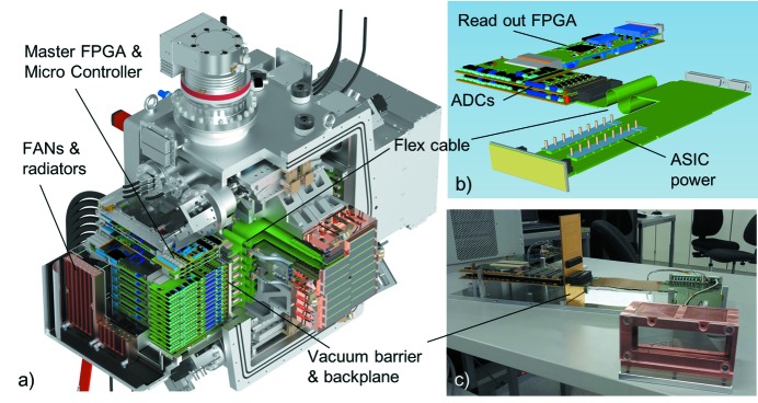
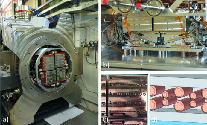
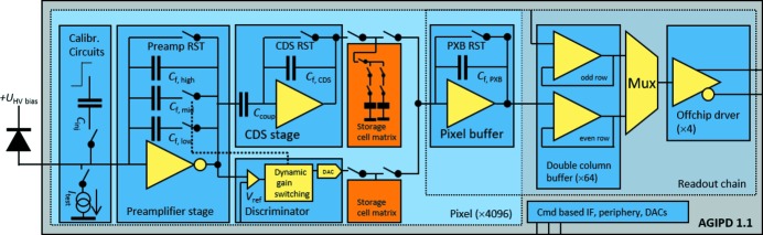
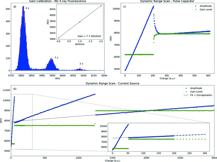
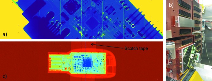
The Adaptive Gain Integrating Pixel Detector (AGIPD) is an X-ray imager, custom designed for the European X-ray Free-Electron Laser (XFEL). It is a fast, low-noise integrating detector, with an adaptive gain amplifier per pixel. This has an equivalent noise of less than 1 keV when detecting single photons and, when switched into another gain state, a dynamic range of more than 104 photons of 12 keV. In burst mode the system is able to store 352 images while running at up to 6.5 MHz, which is compatible with the 4.5 MHz frame rate at the European XFEL. The AGIPD system was installed and commissioned in August 2017, and successfully used for the first experiments at the Single Particles, Clusters and Biomolecules (SPB) experimental station at the European XFEL since September 2017. This paper describes the principal components and performance parameters of the system.
Keywords: AGIPD; European XFEL; X-ray detector.
open access.
Figures
References
-
- Allahgholi, A., Becker, J., Bianco, L., Delfs, A., Gottlicher, P., Graafsma, H., Hirsemann, H., Jack, S., Klyuev, A., Lange, S., Marras, A., Sheviakov, I., Trunk, U., Xia, Q., Zhang, J., Zimmer, M., Dinapoli, R., Greiffenberg, D., Mezza, D., Mozzanica, A., Schmitt, B., Shi, X., Klanner, R., Schwandt, J., Gronewald, M., Kruger, H. & Rah, S. (2014). 2014 IEEE Nucl. Sci. Symp. Med. Imaging Conf. pp. 4799–6097.
-
- Altarelli, M. (2011). Nucl. Instrum. Methods Phys. Res. B, 269, 2845–2849.
-
- Becker, J., Eckstein, D., Klanner, R. & Steinbrück, G. (2010). Nucl. Instrum. Methods Phys. Res. A, 615, 230–236.
-
- Becker, J., Gottlicher, P., Graafsma, H., Hirsemann, H., Jack, S., Klyuev, A., Lange, S., Marras, A., Nilsson, B., Tian, F., Trunk, U., Klanner, R., Schwandt, J., Zhang, J., Dinapoli, R., Greiffenberg, D., Henrich, B., Mozzanica, A., Schmitt, B., Shi, X., Gronewald, M., Karagounis, M. & Kruger, H. (2011). 2011 IEEE Nucl. Sci. Symp. Med. Imaging Conf. pp. 1950–1954.
-
- Blaj, G., Caragiulo, P., Carini, G., Dragone, A., Haller, G., Hart, P., Hasi, J., Herbst, R., Kenney, C., Markovic, B., Nishimura, K., Pines, J., Segal, J., Tamma, C. & Tomada, A. (2016). AIP Conf. Proc. 1741, 040012.
LinkOut - more resources
Full Text Sources
Other Literature Sources
