. 2019 Jan 17;9(1):151.
doi: 10.1038/s41598-018-36379-z.

Autophagy-lysosome pathway alterations and alpha-synuclein up-regulation in the subtype of neuronal ceroid lipofuscinosis, CLN5 disease

Affiliations

Autophagy-lysosome pathway alterations and alpha-synuclein up-regulation in the subtype of neuronal ceroid lipofuscinosis, CLN5 disease

Jessie Adams et al. Sci Rep. .

Abstract

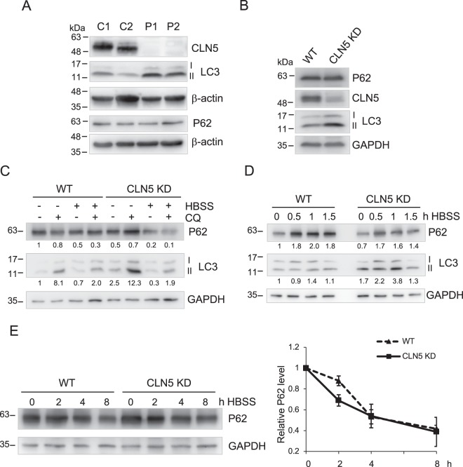
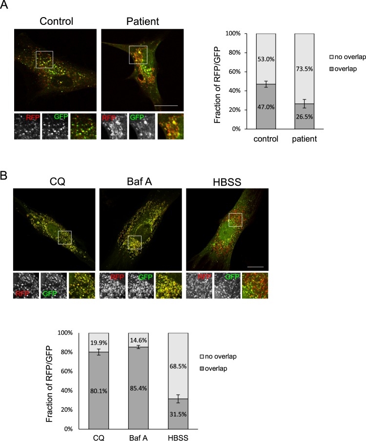
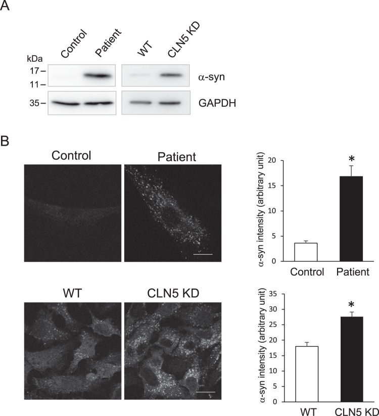
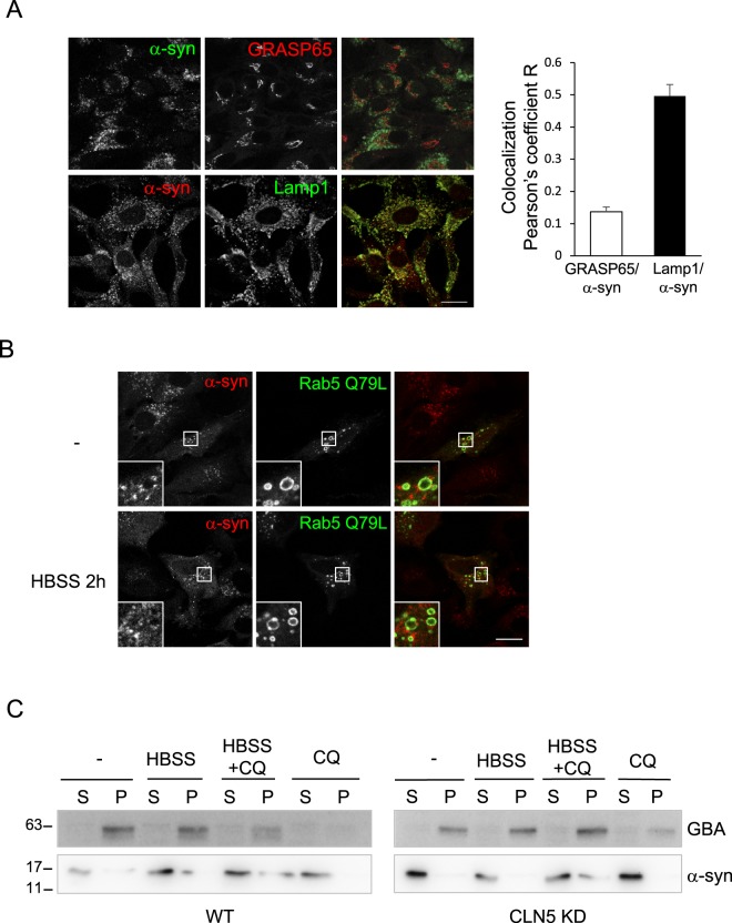
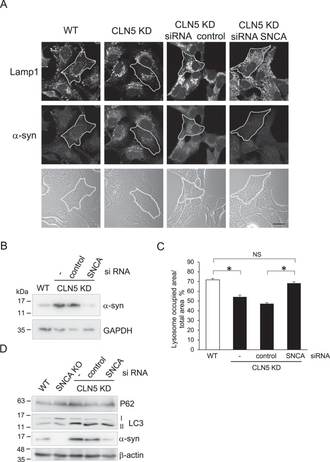
Neuronal ceroid lipofuscinoses (NCLs) are a group of inherited neurodegenerative lysosomal storage disorders. CLN5 deficiency causes a subtype of NCL, referred to as CLN5 disease. CLN5 is a soluble lysosomal protein with an unclear function in the cell. Increased levels of the autophagy marker protein LC3-II have been reported in several subtypes of NCLs. In this report, we examine whether autophagy is altered in CLN5 disease. We found that the basal level of LC3-II was elevated in both CLN5 disease patient fibroblasts and CLN5-deficient HeLa cells. Further analysis using tandem fluorescent mRFP-GFP-LC3 showed the autophagy flux was increased. We found the alpha-synuclein (α-syn) gene SNCA was highly up-regulated in CLN5 disease patient fibroblasts. The aggregated form of α-syn is well known for its role in the pathogenicity of Parkinson's disease. Higher α-syn protein levels confirmed the SNCA up-regulation in both patient cells and CLN5 knockdown HeLa cells. Furthermore, α-syn was localized to the vicinity of lysosomes in CLN5 deficient cells, indicating it may have a lysosome-related function. Intriguingly, knocking down SNCA reversed lysosomal perinuclear clustering caused by CLN5 deficiency. These results suggest α-syn may affect lysosomal clustering in non-neuronal cells, similar to its role in presynaptic vesicles in neurons.


Conflict of interest statement

The authors declare no competing interests.

Figures

Figure 1
Figure 1
Autophagy is enhanced in CLN5-deficient cells. (A) Total lysates of two healthy control fibroblasts (C1 and C2) and two CLN5 disease patient fibroblasts (P1 and P2) were analyzed by immunoblotting. Basal levels of autophagic markers LC3-II and P62 were shown. The CLN5 was absent in patient cells. (B) Total lysates of WT and stable CLN5 KD HeLa cells were analyzed by immunoblotting. The CLN5 was greatly reduced in CLN5 KD cells. Basal levels of autophagic markers LC3-II and P62 were shown. (C) WT and Stable CLN5 KD HeLa cells were treated with CQ, HBSS, or HBSS + CQ for 4 h. Samples were analyzed by immunoblotting. The relative amounts of LC3-II and P62 after normalization with GAPDH are indicated. (D) WT and Stable CLN5 KD HeLa cells were incubated with HBSS for 0, 0.5, 1, 1.5 h. Samples were analyzed by immunoblotting. The relative amounts of LC3-II and P62 after normalization with GAPDH are indicated. (E) WT and Stable CLN5 KD HeLa cells were incubated with HBSS for 0, 2, 4, 8 h in the presence of cycloheximide and bortezomib. Samples were analyzed by immunoblotting. For degradation quantification on the right (N = 3), P62 was normalized with GAPDH signal in each lane. 0 h in each cell line was set as 1. Error bar represents SEM. β-actin and GAPDH were blotted as loading controls. All experiments were repeated at least three times.
Figure 2
Figure 2
More autolysosomes are present in CLN5-deficient patient fibroblasts. (A) Control or CLN5-deficient fibroblasts that had been transfected with the mRFP-GFP-LC3 plasmid for 24 hours were fixed. GFP and RFP signal were visualized using confocal microscopy. The fraction of RFP/GFP signals that was overlapping was analyzed by the Mander’s coefficient using the ImageJ JACoP plugin. In ImageJ, two channels of each image file were first split to two separate images. These two images were then used for plugin JACoP analysis. The threshold of each image was adjusted to reduce the background signals before performing JACoP Mander’s coefficient. n = 15 cells, error bar represents SEM. (B) Control fibroblasts that had been transfected with the mRFP-GFP-LC3 plasmid for 24 hours were treated with CQ, baf A, or HBSS for 2 h prior to fixation. The fraction of RFP/GFP signals that was overlapping was analyzed by the Mander’s coefficient using the ImageJ JACoP plugin. n = 7 cells, error bar represents SEM. All experiments were repeated at least three times. Scale bar: 20 μm.
Figure 3
Figure 3
Higher levels of α-syn were detected in CLN5-deficient cells. (A) Total lysates from control and CLN5 disease patient fibroblasts, as well as WT and CLN5 KD HeLa cells were analyzed by immunoblotting. GAPDH was blotted as a loading control. (B) Control and CLN5 disease patient fibroblasts (top), as well as WT and CLN5 KD HeLa cells (bottom) were immunostained for α-syn. Signal intensity was quantified using ImageJ. Background was subtracted from the total intensity of each cell measured to obtain the correct total cell fluorescence. Data was analyzed using a Student’s t test with two-samples assuming equal variances. *P < 0.0005, n = 5 cells for fibroblasts; *P < 0.0005, n = 20 cells for WT and CLN5 KD HeLa cells, error bar represents SEM. Images were acquired using confocal microscopy. Scale bar: 20 μm. All experiments were repeated at least three times.
Figure 4
Figure 4
α-syn localizes to the vicinity of lysosomes. (A) CLN5 KD HeLa cells were fixed and immunostained for a Golgi marker, GRASP65 (top panel) and a lysosome marker, Lamp1 (bottom panel). The ImageJ JACoP plugin was used to analyze colocalization (Pearson’s coefficient). In ImageJ, two channels of each image file were first split to two separate images. These two images were then used for plugin JACoP analysis. Five images in each set (total 59 cells in GRASP/α-syn set; total 42 cells in Lamp1/α-syn set) were analyzed. Error bar represents SEM. (B) α-syn does not localize to the lumen of lysosomes. CLN5 KD HeLa cells that had been transfected with the EGFP-Rab5 Q79L plasmid for 24 hours were incubated with HBSS for 2 h prior to fixation and immunostained for α-syn. In control (−), cells were left untreated. Cells were fixed and immunostained for CLN5. Images were acquired using confocal microscopy. Scale bar: 20 μm. (C) Subcellular fractionation of α-syn. WT and Stable CLN5 KD HeLa cells were treated with CQ, HBSS, or HBSS + CQ for 2 h. After fractionation, samples were analyzed by immunoblotting. S, soluble; P, pellet. All experiments were repeated at least three times.
Figure 5
Figure 5
Knockdown of α-syn reverses the perinuclear lysosome clustering phenotype found in CLN5 KD HeLa cells. (A) HeLa or CLN5 KD cells were transfected with control or SNCA small interfering RNA (siRNA) for 48 hours prior to fixation. Lysosomes were visualized by immunostaining lamp1. Scale bar: 20 μm. (B) Immunoblot shows efficient knockdown of α-syn expression in CLN5 KD cells. GAPDH was blotted as a loading control. (C) The cellular distribution area of lysosomes was quantified in the conditions indicated. An outline of the cells was marked by tracing the cell border in phase contrast images. The total area of the cell and the area with lamp1 signal in the cell were measured using ImageJ “measure” function. One-way ANOVA followed by the Tukey’s post hoc test was performed *P < 0.05, n = 25, error bar represents SEM. (D) Immunoblotting of WT, stable SNCA knock-out cell (SNCA KO), and siRNA of stable CLN5 KD as indicated. β-actin was blotted as a loading control. All experiments were repeated at least three times.

References

    1. Bennett MJ, Hofmann SL. The neuronal ceroid-lipofuscinoses (Batten disease): a new class of lysosomal storage diseases. J Inherit Metab Dis. 1999;22:535–544. doi: 10.1023/A:1005564509027. - DOI - PubMed
    1. Haltia M. The neuronal ceroid-lipofuscinoses: from past to present. Biochim Biophys Acta. 2006;1762:850–856. doi: 10.1016/j.bbadis.2006.06.010. - DOI - PubMed
    1. Williams RE, Mole SE. New nomenclature and classification scheme for the neuronal ceroid lipofuscinoses. Neurology. 2012;79:183–191. doi: 10.1212/WNL.0b013e31825f0547. - DOI - PubMed
    1. Palmer DN, et al. Mitochondrial ATP synthase subunit c storage in the ceroid-lipofuscinoses (Batten disease) Am J Med Genet. 1992;42:561–567. doi: 10.1002/ajmg.1320420428. - DOI - PubMed
    1. Tyynela J, Palmer DN, Baumann M, Haltia M. Storage of saposins A and D in infantile neuronal ceroid-lipofuscinosis. FEBS Lett. 1993;330:8–12. doi: 10.1016/0014-5793(93)80908-D. - DOI - PubMed

Publication types