. 2019 Jan 17;9(1):195.
doi: 10.1038/s41598-018-36808-z.

The Phenolic compound Kaempferol overcomes 5-fluorouracil resistance in human resistant LS174 colon cancer cells

Affiliations

The Phenolic compound Kaempferol overcomes 5-fluorouracil resistance in human resistant LS174 colon cancer cells

Ichrak Riahi-Chebbi et al. Sci Rep. .

Abstract

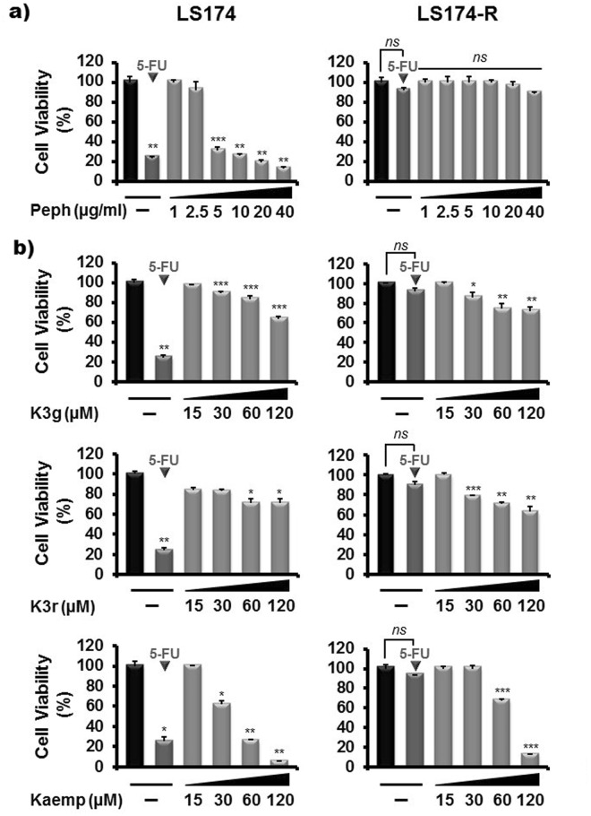
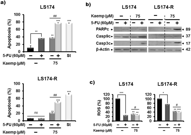
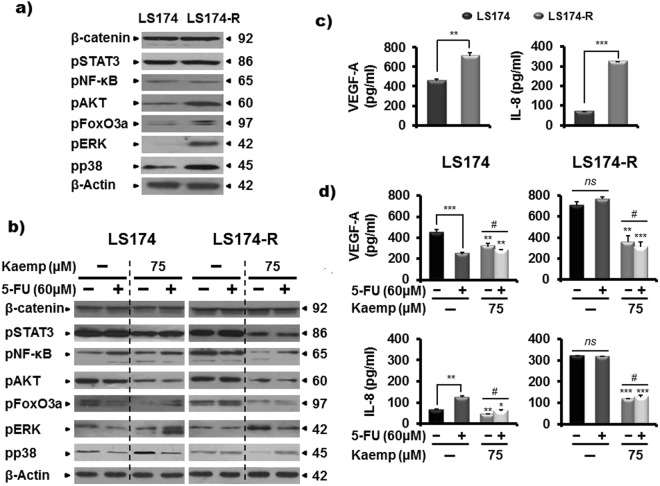
Resistance to 5-Fluorouracil chemotherapy is a major cause of therapeutic failure in colon cancer cure. Development of combined therapies constitutes an effective strategy to inhibit cancer cells and prevent the emergence of drug resistance. For this purpose, we investigated the anti-tumoral effect of thirteen phenolic compounds, from the Tunisian quince Cydonia oblonga Miller, alone or combined to 5-FU, on the human 5-FU-resistant LS174-R colon cancer cells in comparison to parental cells. Our results showed that only Kaempferol was able to chemo-sensitize 5-FU-resistant LS174-R cells. This phenolic compound combined with 5-FU exerted synergistic inhibitory effect on cell viability. This combination enhanced the apoptosis and induced cell cycle arrest of both chemo-resistant and sensitive cells through impacting the expression levels of different cellular effectors. Kaempferol also blocked the production of reactive oxygen species (ROS) and modulated the expression of JAK/STAT3, MAPK, PI3K/AKT and NF-κB. In silico docking analysis suggested that the potent anti-tumoral effect of Kaempferol, compared to its two analogs (Kaempferol 3-O-glucoside and Kampferol 3-O-rutinoside), can be explained by the absence of glucosyl groups. Overall, our data propose Kaempferol as a potential chemotherapeutic agent to be used alone or in combination with 5-FU to overcome colon cancer drug resistance.


Conflict of interest statement

The authors declare no competing interests.

Figures

Figure 1
Figure 1
Characteristics of 5-FU-resistant LS174-R cells in comparison to the parental ones. (a) Representative microscopic images of both sensitive and 5-FU resistant cancer cells photographed under phase contrast microscope. (b) Proliferation rate of parental and 5-FU resistant cells for 24 h, 48 h and 72 h was determined by MTT assay. (c) Colony-forming capacity of parental and 5-FU-resistant cells was measured using clonogenic survival assay. 2000 viable cells from each group were cultured in six-well plates for additional 10 days. Colonies were stained with crystal violet and each assay was photographed. (d) Representative microscopy images of colon cancer cells in 3D cultures (x10). Both colon cancer cells were analyzed for spheroid formation capacity in ultra-low attachment (ULA) round bottom 96-well plates coated with agarose. (e) Cell cycle analysis of 5-FU-resistant cells compared to parental cells by flow cytometry using propidium iodide assay. (f) Protein expression of different cellular effectors was analyzed in both parental LS174 and 5-FU resistant LS174-R cells by western blot using specific antibodies. β-actin was used as a reference protein for equal loading. (g) Sensitive LS174 and resistant LS174-R cells were treated with different concentrations of 5-FU. The IC50 (50% inhibitory concentration) values were calculated at 72 h time post treatment with MTT assay. Results were normalized to each control in percentage and represented as mean ± SE of three independent experiments, each performed at least in triplicate. *p < 0.05, **p < 0.01, ***p < 0.005 and ns: non significant.
Figure 2
Figure 2
Kaempferol overcomes 5-FU resistance in LS174-R cancer cells. Parental LS174 cells and resistant LS174-R cells were cultured in 96-well plates and treated with increasing concentrations of (a) Cydonia oblonga Miller peel polyphenolic extract (Peph) (1–40 µg/ml) and (b) Kaempferol and its analogs, Kaempferol 3-O-glucoside (K3g) and Kaempferol 3-O-rutinoside (K3r), (15–120 µM) for 72 h. Cell viability was measured by MTT assay. The absorbance was measured at 540 nm. Results were normalized to each control in percentage and represented as mean ± SE of three independent experiments. *p < 0.05, **p < 0.01, ***p < 0.005 when compared to their respective CN and ns: non significant.
Figure 3
Figure 3
Kaempferol chemo-sensitizes resistant colon cancer cells to 5-FU chemotherapy. (a) Parental LS174 cells and resistant LS174-R cells were cultured in 96-well plates and treated with a concentration range from 15 µM to 120 µM of Kaempferol alone, 5-FU (60 µM) alone and the combination of both for 72 h. Cell viability was measured by MTT assay. The absorbance was measured at 540 nm. Assays were performed in triplicate. (b) Kaempferol effect on parental LS174 cells and resistant LS174-R cells colony-forming capacity was measured using clonogenic survival assay. Colon cancer cells were treated with vehicle (control) or Kaempferol (15 and 75 µM) combined or not to 60 µM of 5-FU for 72 h. After removal of the medium, 2000 viable cells from each group were cultured in six-well plates for additional 10 days. Colonies were stained with crystal violet and each assay was photographed. (c) The number of colonies was analyzed and scored by CFU scope quantification software. Results are expressed as the number of colony forming cells per well in percentage and normalized to control (vehicle, considered to represent 100%). Graphs are represented as mean ± SD of three independent experiments. *p < 0.05, **p < 0.01 and ***p < 0.005 when compared to their respective controls. #p < 0.05, ##p < 0.01 (Kaempferol + 5-FU groups vs Keampferol groups).
Figure 4
Figure 4
Kaempferol induces cell cycle arrest of sensitive and 5-FU-resistant cancer cells. (a) Cell cycle phase distribution of cancer cells cultured in the absence (mock) and the presence of Kaempferol (75 µM) alone or combined to 60 µM of 5-FU for 72 h were analyzed by flow cytometry using propidium iodide assay. Results were represented as mean ± SE of three independent experiments. *p < 0.05, **p < 0.01, ***p < 0.005 when compared to their respective CN, #p < 0.05, ##p < 0.01 (Kaempferol + 5-FU groups vs Keampferol groups) and ns: non significant. Cell cycle-related proteins were analyzed by western blotting in (b) 5-FU resistant cells versus parental cells and (c) after cell treatments with Kaempferol, 5-FU, and the combination of both for 72 h. β-actin was used as a loading control. The data shown are representative of three independent experiments.
Figure 5
Figure 5
Kaempferol chemosensitizes 5-FU-resistant cancer cells to apoptosis and reduces the ROS production. (a) Apoptosis detection in mock and treated-cells with Kaempferol alone or combined to 5-FU for 72 h by flow cytometry analysis using annexin-V/7-AAD staining. Staurosporin (2 µM, St) was used as a positive control of apoptosis. Results were represented as mean ± SE of three independent experiments. *p < 0.05, **p < 0.01, ***p < 0.005 when compared to their respective CN, #p < 0.05, ##p < 0.01 (Kaempferol + 5-FU groups vs Keampferol groups) and ns: non significant. (b) Western blot analysis of some apoptosis-related proteins in both parental LS174 and 5-FU resistant LS174-R cells after 72 h of treatment with Kaempferol, 5-FU and the combination of both. β-actin was used as a reference protein for equal loading. The data shown are representative of three independent experiments. (c) Histograms analysis of ROS production measured with CMH2DCFDA staining after 72 h of treatment with Kaempefrol, 5-FU and the combination of both in parental LS174 and 5-FU-resistant LS174-R cells. Detection of ROS was related to the quantity of subsequent oxidation leading emitting fluorescence. Data are reported as the means ± S.E.M of three independent experiments. *p < 0.05, **p < 0.01, *** p < 0.005 with respect to mock-treated controls; #p < 0.05, ##p < 0.01 (Kaempferol + 5-FU groups vs Keampferol groups).
Figure 6
Figure 6
Kaempferol modulates survival signaling pathways in both sensitive and resistant colon cancer cells and reduces the production of the two angiogenic factors, VEGF-A and IL-8 in 5-FU-refractive LS174-R cells. (a) Different cellular effectors were monitored in chemo-resistant LS174-R cells and sensitive LS174 cells by western blot using specific antibodies. β-actin was used as a loading control. (b) 5-FU-sensitive and resistant cancer cells were treated with Kaempferol, 5-FU, and the combination of both for 72 h. Protein extracts (30 µg) from whole cell lysates were analysed by western blotting using specific antibodies. β-actin was used as a reference protein for equal loading. One representative experiment of three independent ones was shown. (c) VEGF and IL-8 secretion were determined by human VEGF and IL-8 ELISA Kit in both control sensitive and 5-FU-resistant LS174-R cells. (d) Supernatants from parental LS174 and 5-FU resistant LS174-R cells cultured in the absence (vehicle) or presence of Kaempferol (75 µM) combined or not to 60 µM of 5-FU were collected and analyzed by Human VEGF and IL-8 specific ELISA. Results are reported as the mean ± SE of three independent experiments each run in triplicate (*p < 0.05, **p < 0.01, *** p < 0.005; #p < 0.05, ##p < 0.01 (Kaempferol + 5-FU groups vs Keampferol groups and ns: non significant). The data were corrected to the cell number.
Figure 7
Figure 7
Kaempferol modulates the expression levels of enzymes involved in 5-FU metabolism. (a) The amounts of mRNA transcripts of five genes, Thymidylate synthase (TS), Thymidine kinase (TK), Dihydrofolate reductase (DHFR), Folylpolyglutamate synthetase (FPGS) and Dihydropyrimidine dehydrogenase (DPD), involved in 5-FU metabolism were quantified by real time PCR in control sensitive and 5-FU-resitant cells and (b) in LS174 and LS174-R cells cultured in the absence (mock) and the presence of Kaempferol (75 µM) and/or 5-FU (60 µM) for 72 h. The values are normalized to GAPDH gene and the control value was taken as 1. Results are reported as the mean ± SE of three independent experiments each run in duplicate (*p < 0.05, **p < 0.01, *** p < 0.005; #p < 0.05, ##p < 0.01 (Kaempferol + 5-FU treatments vs Keampferol groups), ns: non significant) (c) TS and TK protein levels were assessed by western blotting in both control 5-FU-sensitive LS174 and resistant LS174-R cells and in (d) colon cancer cells after treatment with vehicle (mock), Kaempferol (75 µM), 5-FU (60 µM), and the combination of both for 72 h. β-actin was used as a reference protein for equal loading.
Figure 8
Figure 8
Computational interaction study of Kaempferol and its analogs with putative human cell proteins. (a) Interaction network of the different proteins from which expression was evaluated by the western blot assay (represented by yellow nodes). Other proteins (blue nodes) emerged from the analysis as connectors of the input proteins. The degree of centrality is proportional to the node radius. (b) Docking of Kaempferol (yellow sticks) with PIM1. The first panel shows the good superposition of Kaempferol with Fisetin structure (Green sticks). The second and the third panel show the docking complexes of Kaempferol 3-O-rutinoside (K3r) and Kaempferol 3-O-glucoside (K3g) with PIM1 respectively, compared to the Kaempferol. (c) Docking solutions of Kaempferol (Yellow sticks) with the ATP binding site in MAPK14 (p38). The two other panels show the position of Kaempferol in the interaction site in comparison to the retained complexes for K3g and K3r. Unlike Kaempferol, these two ligands cannot attain the depth of the binding pocket.

References

    1. Murphy, N. et al. Heterogeneity of Colorectal Cancer Risk Factors by Anatomical Subsite in 10 European Countries: A Multinational Cohort Study. Clin Gastroenterol Hepatol, 10.1016/j.cgh.2018.07.030 (2018). - PMC - PubMed
    1. Bray, F. et al. Global Cancer Statistics 2018: GLOBOCAN Estimates of Incidence and Mortality Worldwide for 36 Cancers in 185 Countries. CA Cancer J Clin, 10.3322/caac.21492 (2018). - PubMed
    1. Landre T, et al. Doublet chemotherapy vs. single-agent therapy with 5FU in elderly patients with metastatic colorectal cancer. a meta-analysis. Int J Colorectal Dis. 2015;30:1305–1310. doi: 10.1007/s00384-015-2296-5. - DOI - PubMed
    1. Mármol I, Sánchez-de-Diego C, Pradilla Dieste A, Cerrada E, Rodriguez Yoldi M. Colorectal Carcinoma: A General Overview and Future Perspectives in Colorectal Cancer. Int J Mol Sci. 2017;18:197. doi: 10.3390/ijms18010197. - DOI - PMC - PubMed
    1. Webber EM, Kauffman TL, O’Connor E, Goddard KA. Systematic review of the predictive effect of MSI status in colorectal cancer patients undergoing 5FU-based chemotherapy. BMC Cancer. 2015;15:156. doi: 10.1186/s12885-015-1093-4. - DOI - PMC - PubMed

Publication types

MeSH terms