Review
. 2019 Mar;20(3):175-193.
doi: 10.1038/s41580-018-0089-8.

Regulation of apoptosis in health and disease: the balancing act of BCL-2 family proteins

Affiliations
Review

Regulation of apoptosis in health and disease: the balancing act of BCL-2 family proteins

Rumani Singh et al. Nat Rev Mol Cell Biol. 2019 Mar.

Abstract

The loss of vital cells within healthy tissues contributes to the development, progression and treatment outcomes of many human disorders, including neurological and infectious diseases as well as environmental and medical toxicities. Conversely, the abnormal survival and accumulation of damaged or superfluous cells drive prominent human pathologies such as cancers and autoimmune diseases. Apoptosis is an evolutionarily conserved cell death pathway that is responsible for the programmed culling of cells during normal eukaryotic development and maintenance of organismal homeostasis. This pathway is controlled by the BCL-2 family of proteins, which contains both pro-apoptotic and pro-survival members that balance the decision between cellular life and death. Recent insights into the dynamic interactions between BCL-2 family proteins and how they control apoptotic cell death in healthy and diseased cells have uncovered novel opportunities for therapeutic intervention. Importantly, the development of both positive and negative small-molecule modulators of apoptosis is now enabling researchers to translate the discoveries that have been made in the laboratory into clinical practice to positively impact human health.


Figures

Figure 1:
Figure 1:. The mitochondrial apoptosis pathway.
To initiate apoptosis, cellular stress or damage signals [1] unleash pro-apoptotic proteins (BH3-only ‘activators’ of apoptosis) via their upregulation (BIM or PUMA) or cleavage (BID cleaved to form truncated tBID) [2], which can either be bound and sequestered by pro-survival proteins such as BCL-2, BCL-XL or MCL1 [3] or, when these pro-survival proteins are saturated or absent, can activate BAX and/or BAK [4]. Activated BAX or BAK oligomerize and form pores to cause mitochondrial outer membrane permeabilization (MOMP), resulting in the release of apoptogenic molecules including SMAC, OMI and cytochrome c from the mitochondrial intermembrane space. Cytochrome c binds APAF1 in the cytosol to form the apoptosome (5), which serves as a platform for the activation of caspase 9, which then goes on to activate the effector caspases 3 and 7 (6) to dismantle the cell and prepare it for phagocytosis. Caspase activation can be blocked by XIAP (7), which in turn is inhibited by the released SMAC and OMI proteins from mitochondria (7). Upstream damage or stress signalling can also activate BH3-only ‘sensitizer’ proteins that don’t efficiently activate BAX and BAK but inhibit the activity of pro-survival BCL-2 family proteins to release any sequestered BH3-only activators, which trigger MOMP (8). BH3 mimetics are a novel class of agents that are able to sensitize cells to apoptosis by blocking the activity of pro-survival BCL-2 family proteins (9).
Figure 2:
Figure 2:. Interactions among BCL-2 family proteins and apoptotic priming.
(a) BCL-2 family proteins interact in several key ways. BH3-only ‘activator’ proteins (BIM, tBID and to a lesser extent PUMA (dashed lines)) can activate BAX or BAK to result in mitochondrial permeabilization (tBID most efficiently activates BAK, whereas BIM shows stronger affinity for BAX). However, pro-survival proteins can also bind and sequester the activator BH3-only proteins via Mode 1 inhibition. BH3-only ‘sensitizer’ proteins can bind and inactivate specific pro-survival proteins, which would result in the release of any BH3-only activators that are actively being sequestered. Finally, pro-survival proteins can also bind and sequester BAX or BAK directly via Mode 2 inhibition, preventing their oligomerization. Damage and stress-induced signalling pathways trigger apoptosis via distinct mechanisms involving modulation of the expression levels or activity of BCL-2 family proteins. The table below is a graphic depiction of the different interactions and their affinities established between BCL-2 family proteins. These different interactions result in differential regulation of apoptosis, depending on expression levels of the different components. In addition, BH3 mimetics of sensitizer proteins can be used to further modify the BCL-2 protein interactome. (b) Cells can exhibit widely differing susceptibility to apoptosis. Cells which are primed for apoptosis express high levels of pro-apoptotic activator proteins at a basal state and even a mild stress, which causes a modest increase in the expression of pro-apoptotic factors, will trigger mitochondrial outer membrane permeabilization (MOMP). Unprimed cells can activate apoptosis, but require more damage or stress to overwhelm pro-survival proteins that are not actively sequestering pro-apoptotic molecules. Finally, cells that are apoptosis refractory do not express sufficient levels of the pro-apoptotic pore-forming proteins BAX or BAK and are unable to trigger MOMP even under severe stress.
Figure 3:
Figure 3:. Apoptosis and apoptotic priming in physiology.
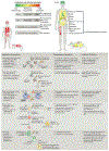
Apoptosis is differently and dynamically regulated across mammalian lifespan. Tissues that are highly proliferative or have the potential to become highly proliferative (developing tissues, adult haematopoietic system) are typically primed for apoptosis (red). High apoptotic priming in these tissues makes them highly sensitive to various insults. Tissues that are largely post-mitotic are apoptosis refractory (green), whereas tissues that are characterized as unprimed (such as gastrointestinal system and lungs of adults) (yellow) typically contain highly heterogeneous cell types that may differ in apoptosis sensitivity at a cell by cell level. The level of priming within cells or tissues is dependent on the expression of BCL-2 family proteins, with the strongest determinants being expression of the pro-apoptotic, pore-forming molecules BAX and BAK. Pro-survival and sensitizer BH3-only proteins can be expressed at variable levels and are not as clearly associated with a specific level of priming. Changes in the levels of BCL-2 proteins will, inevitably, impose changes on apoptotic susceptibility, leading to increased or insufficient apoptosis, which can result in pathology. HSC, haematopoietic stem cell; IR, ionizing radiation.
Figure 4:
Figure 4:. Apoptotic dependencies in cancer cells and their responsiveness to therapy.
(a) During the process of neoplastic transformation, oncogene-driven abnormal growth signals and cell-cycle checkpoint violation [G] leads to cellular stress and upregulation of pro-apoptotic proteins. This upregulation results in higher apoptotic priming of malignant cells at the basal state than normal cells. Because healthy adult tissues are mostly refractory to apoptosis or unprimed (see also Fig. 3), this increase in apoptotic priming of aberrant cells can be exploited therapeutically using BH3 mimetics, in particular when combined with standard anti-cancer therapies such as radiation or chemotherapy. (b) Haematopoietic cells are naturally highly primed for apoptosis. Hence, oncogenic stress and resulting upregulation of pro-apoptotic factors frequently results in the removal of pre-malignant cells derived from this lineage (middle arrow). Several mechanisms associated with BCL-2 protein deregulation, including BIM mutations (1), downregulation of BAX (2) or BIM (3) and upregulation of BCL-2 or MCL1 (4) support the emergence of haematopoietic cancers, which depend on these mechanisms for their survival. Notably, these mechanisms are mainly employed to keep malignant cells alive and haematopoietic cancers typically remain primed for apoptosis and hence are susceptible to therapy and respond well to treatments with BH3 mimetics. However, certain mechanisms, including mutations of sensitizer proteins (1) and downregulation of mitochondrial outer membrane permeabilization (MOMP) pore-forming components (2) yield cells that are unprimed or even apoptosis refractory, and hence resistant to therapies. Solid tumours can employ similar mechanisms to boost their survival, but these dependencies are much less pronounced than in haematopoietic cancers. Nevertheless, these mechanisms may underlie the development of resistance to treatment and disease relapse.
None

References

    1. Rathmell JC & Thompson CB Pathways of apoptosis in lymphocyte development, homeostasis, and disease. Cell 109, S97–107 (2002). - PubMed
    1. Sedger LM et al. Extreme lymphoproliferative disease and fatal autoimmune thrombocytopenia in FasL and TRAIL double-deficient mice. Blood 115, 3258–68 (2010). - PMC - PubMed
    1. Lamhamedi-Cherradi SE, Zheng SJ, Maguschak KA, Peschon J & Chen YH Defective thymocyte apoptosis and accelerated autoimmune diseases in TRAIL−/− mice. Nat. Immunol 4, 255–260 (2003). - PubMed
    1. Su JH, Deng G & Cotman CW Bax Protein Expression Is Increased in Alzheimer’s Brain: Correlations with DNA Damage, Bcl-2 Expression, and Brain Pathology. J. Neuropathol. Exp. Neurol 56, 86–93 (1997). - PubMed
    1. Lu T et al. REST and stress resistance in ageing and Alzheimer’s disease. Nature 507, 448–54 (2014). - PMC - PubMed

Publication types

Substances