

NANOGP8 expression regulates gastric cancer cell progression by transactivating DBC1 in gastric cancer MKN-45 cells
- PMID: 30655801
- PMCID: PMC6313168
- DOI: 10.3892/ol.2018.9595
NANOGP8 expression regulates gastric cancer cell progression by transactivating DBC1 in gastric cancer MKN-45 cells
Abstract
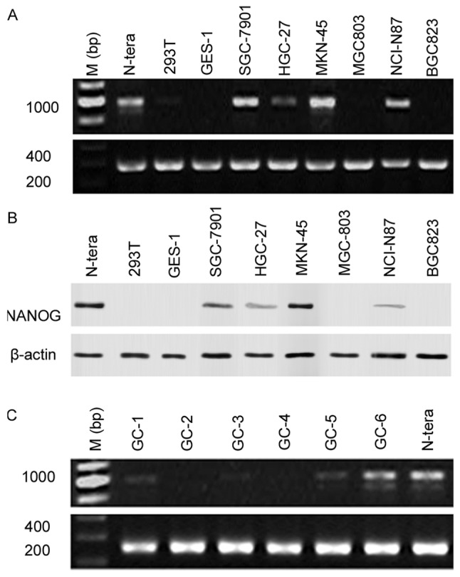
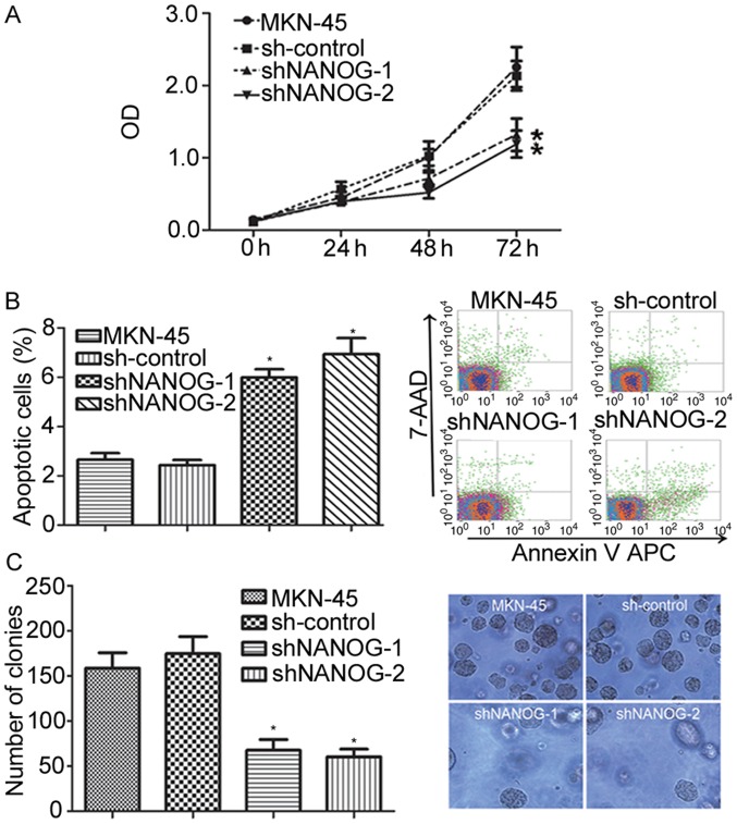
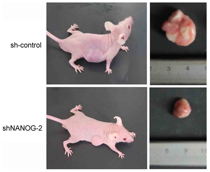
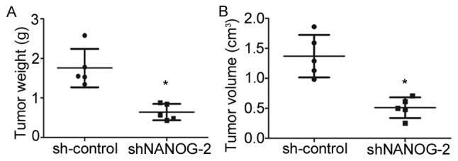
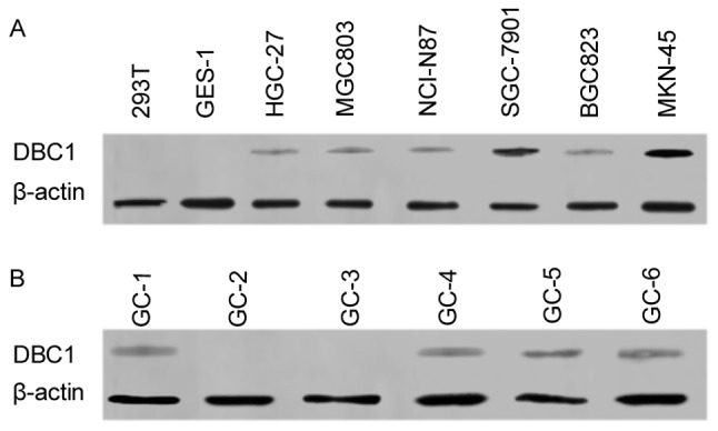
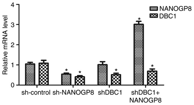
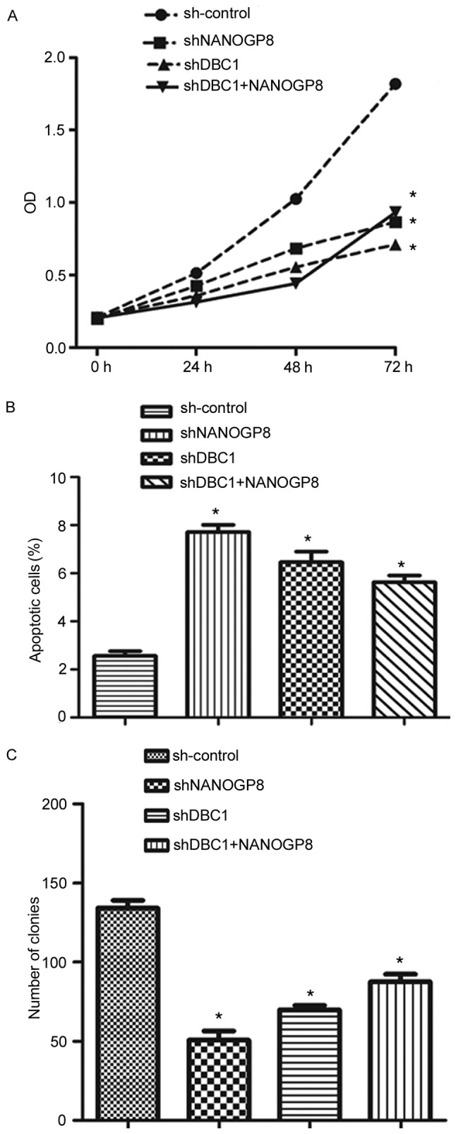
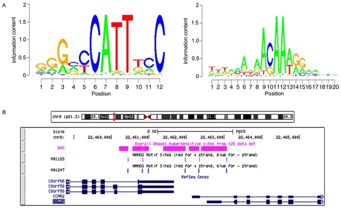
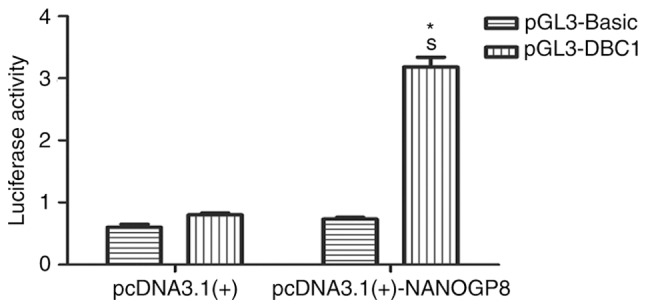
NANOGP8 is one of the NANOG pseudogenes and is expressed together with NANOG in multiple tumor tissues and cell lines. The biological functions of NANOGP8 in progression of gastric cancer are unclear. In the present study, the role of NANOGP8 was investigated in gastric cancer cells. The gathered data demonstrated that NANOG expression in both mRNA and protein was elevated in gastric cancer cell lines relative to a normal gastric epithelial cell line. Downregulation of NANOGP8 inhibited cell proliferation and increased apoptosis in human gastric carcinoma cell lines. Furthermore, silencing of NANOGP8 suppressed tumor growth in vivo. Interestingly, it was identified that deleted in breast cancer 1 (DBC1) expression was also markedly downregulated following NANOGP8 knockdown. DNA microarray and dual-luciferase assays further indicated that NANOGP8 may bind to the DBC1 promoter region and regulate DBC1 expression. Therefore, the gathered data provided evidence that NANOGP8 contributes to progression of gastric cancer via DBC1 and may have potential translational significance.
Keywords: apoptosis; gastric cancer; proliferation.
Figures
Similar articles
-
NANOGP8 is the key regulator of stemness, EMT, Wnt pathway, chemoresistance, and other malignant phenotypes in gastric cancer cells.PLoS One. 2018 Apr 24;13(4):e0192436. doi: 10.1371/journal.pone.0192436. eCollection 2018. PLoS One. 2018. PMID: 29689047 Free PMC article.
-
Oncogenic NanogP8 expression regulates cell proliferation and migration through the Akt/mTOR signaling pathway in human gastric cancer - SGC-7901cell line.Onco Targets Ther. 2016 Aug 9;9:4859-66. doi: 10.2147/OTT.S97861. eCollection 2016. Onco Targets Ther. 2016. PMID: 27563247 Free PMC article.
-
The human pluripotency gene NANOG/NANOGP8 is expressed in gastric cancer and associated with tumor development.Oncol Lett. 2010 May;1(3):457-463. doi: 10.3892/ol_00000081. Epub 2010 May 1. Oncol Lett. 2010. PMID: 22966326 Free PMC article.
-
[The expression of retrogene Nanogp8 is related to the biological characteristics of human SGC-7901 gastric cancer cells].Xi Bao Yu Fen Zi Mian Yi Xue Za Zhi. 2015 Jun;31(6):763-8. Xi Bao Yu Fen Zi Mian Yi Xue Za Zhi. 2015. PMID: 26062418 Chinese.
-
Advances on the role of the deleted in breast cancer (DBC1) in cancer and autoimmune diseases.J Leukoc Biol. 2021 Feb;109(2):449-454. doi: 10.1002/JLB.6MR0320-086R. Epub 2020 Apr 26. J Leukoc Biol. 2021. PMID: 32337788 Review.
Cited by
-
Pseudogene-Derived lncRNAs and Their miRNA Sponging Mechanism in Human Cancer.Front Cell Dev Biol. 2020 Feb 28;8:85. doi: 10.3389/fcell.2020.00085. eCollection 2020. Front Cell Dev Biol. 2020. PMID: 32185172 Free PMC article. Review.
-
Pseudogenes as Biomarkers and Therapeutic Targets in Human Cancers.Methods Mol Biol. 2021;2324:319-337. doi: 10.1007/978-1-0716-1503-4_20. Methods Mol Biol. 2021. PMID: 34165724 Review.
-
A Restriction Endonuclease-Based Assay to Distinguish NANOGP8 Retrogene from Parental NANOG.Methods Mol Biol. 2021;2324:255-262. doi: 10.1007/978-1-0716-1503-4_16. Methods Mol Biol. 2021. PMID: 34165720
-
Pseudogene DUXAP8 Promotes Cell Proliferation and Migration of Hepatocellular Carcinoma by Sponging MiR-490-5p to Induce BUB1 Expression.Front Genet. 2020 Jul 22;11:666. doi: 10.3389/fgene.2020.00666. eCollection 2020. Front Genet. 2020. PMID: 32849765 Free PMC article.
-
GENCODE Pseudogenes.Methods Mol Biol. 2021;2324:67-82. doi: 10.1007/978-1-0716-1503-4_5. Methods Mol Biol. 2021. PMID: 34165709
References
LinkOut - more resources
Full Text Sources
Research Materials