. 2019 Jan;17(1):603-615.
doi: 10.3892/ol.2018.9629. Epub 2018 Oct 29.

Synergistic action of microwave-induced mild hyperthermia and paclitaxel in inducing apoptosis in the human breast cancer cell line MCF-7

Affiliations

Synergistic action of microwave-induced mild hyperthermia and paclitaxel in inducing apoptosis in the human breast cancer cell line MCF-7

Sina Liao et al. Oncol Lett. 2019 Jan.

Abstract

Microwave mild hyperthermia and paclitaxel have been reported to be involved in variety of solid tumors. However, rare related researches have been accomplished via directly killing tumor cells using thermochemotherapy. In order to clarify the potential synergy between microwave-induced hyperthermia at temperatures <41°C and paclitaxel chemotherapy for inhibiting the growth of the human breast cancer cell line MCF-7, an MTT assay was used. The MCF-7 cells cultured in vitro were treated with paclitaxel alone, treated with microwave-induced hyperthermia for 2 h alone (at 40, 40.5 or 41°C), or treated with a combination of paclitaxel and 2 h of hyperthermia (at 40, 40.5 or 41°C). Flow cytometry was used to determine the cell apoptosis rate and it was demonstrated that paclitaxel decreased cell viability in a dose-dependent manner. Alone, hyperthermia for 2 h at 41°C induced apoptosis in MCF-7 cells, to a greater extent compared with hyperthermia for 2 h at 40.0 or 40.5°C (P<0.05). Together, paclitaxel and 2 h of hyperthermia at 40.5°C induced significantly increased apoptosis compared with either treatment alone (P<0.05). Increasing the temperature to 41°C in combination with paclitaxel increased the apoptotic ratio from 12.21±1.02% to 16.36±2.39%. The apoptotic ratio correlated positively with hyperthermia temperature and duration following hyperthermia, as did the synergistic effect obtained by combining hyperthermia and paclitaxel. Notably, the combination of 5 µg/ml paclitaxel and 2 h of hyperthermia at 40°C enhanced MCF-7 cell proliferation. Mild hyperthermia may exert anti-tumor effects by inducing apoptosis, and combining hyperthermia with paclitaxel synergistically induces apoptosis. Paclitaxel dose and hyperthermia temperature require careful optimization, as low-dose paclitaxel combined with hyperthermia at an insufficient temperature may enhance breast cancer proliferation.

Keywords: apoptosis; combined treatment; human breast cancer cell line MCF-7; microwave mild hyperthermia; paclitaxel.


Figures
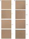
Figure 1.
Figure 1.
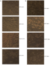
Photomicrographs displaying the effects of mild hyperthermia (37°C, 40°C, 40.5°C and 41°C) on MCF-7 growth at (A) 24 h post-exposure or (B) 48 h post-exposure. Obvious apoptotic cell morphology was observed at 48 h following hyperthermia at 41°C. Magnification, ×100.
Figure 2.
Figure 2.
Effects of mild hyperthermia on the apoptotic ratio of MCF-7 cells. Flow cytometry of MCF-7 cells using an Annexin V-FITC/PI cell apoptosis staining kit. (A) 24 h following hyperthermia or (B) 48 h following hyperthermia. (C) The apoptotic ratio (% apoptotic cells) 24 and 48 h following hyperthermia. **P<0.01 vs. 48 h (41°C); *P<0.05 vs. control group (37°C); #P<0.05 vs. 40.5°C; &P<0.05 vs. 40°C. FITC, fluorescein isothiocyanate; PI, propidium iodide.
Figure 2.
Figure 2.
Effects of mild hyperthermia on the apoptotic ratio of MCF-7 cells. Flow cytometry of MCF-7 cells using an Annexin V-FITC/PI cell apoptosis staining kit. (A) 24 h following hyperthermia or (B) 48 h following hyperthermia. (C) The apoptotic ratio (% apoptotic cells) 24 and 48 h following hyperthermia. **P<0.01 vs. 48 h (41°C); *P<0.05 vs. control group (37°C); #P<0.05 vs. 40.5°C; &P<0.05 vs. 40°C. FITC, fluorescein isothiocyanate; PI, propidium iodide.
Figure 3.
Figure 3.
Anti-proliferative effect of paclitaxel on MCF-7 cells. Cultures were incubated with increasing paclitaxel concentrations, and proliferation was measured using the MTT Cell Proliferation Assay kit. Absorbance at 570 nm was determined using an enzyme-linked immunosorbent assay. IC10 and IC20 were determined to be, 5 and 10 µg/ml, respectively. IC, inhibitory concentration.
Figure 4.
Figure 4.
Photomicrographs displaying the effects of the combination of paclitaxel and mild hyperthermia on MCF-7 cells. (A) MCF-7 cells 24 h post-treatment with 5 µg/ml of paclitaxel and exposure to mild hyperthermia. (B) MCF-7 cells 48 h post-treatment with 5 µg/ml paclitaxel and exposure to mild hyperthermia. (C) MCF-7 cells 24 h post-treatment with 10 µg/ml of paclitaxel and exposure to mild hyperthermia. (D) MCF-7 cells 48 h post-treatment with 10 µg/ml of paclitaxel and exposure to mild hyperthermia. Cultures exposed to hyperthermia at 40°C displayed reduced apoptosis compared with cultures treated with 5 µg/ml paclitaxel alone, while cultures treated with hyperthermia at 40.5 or 41°C displayed greater numbers of dead cells (due to apoptosis or necrosis) compared with cultures treated with 5 or 10 µg/ml or paclitaxel alone. The numbers of dead cells increased with longer time after treatment. The images of 37°C 24 h and 37°C 48 h are the results of paclitaxel (5 or 10 µg/ml) alone. Magnification, ×100.
Figure 4.
Figure 4.
Photomicrographs displaying the effects of the combination of paclitaxel and mild hyperthermia on MCF-7 cells. (A) MCF-7 cells 24 h post-treatment with 5 µg/ml of paclitaxel and exposure to mild hyperthermia. (B) MCF-7 cells 48 h post-treatment with 5 µg/ml paclitaxel and exposure to mild hyperthermia. (C) MCF-7 cells 24 h post-treatment with 10 µg/ml of paclitaxel and exposure to mild hyperthermia. (D) MCF-7 cells 48 h post-treatment with 10 µg/ml of paclitaxel and exposure to mild hyperthermia. Cultures exposed to hyperthermia at 40°C displayed reduced apoptosis compared with cultures treated with 5 µg/ml paclitaxel alone, while cultures treated with hyperthermia at 40.5 or 41°C displayed greater numbers of dead cells (due to apoptosis or necrosis) compared with cultures treated with 5 or 10 µg/ml or paclitaxel alone. The numbers of dead cells increased with longer time after treatment. The images of 37°C 24 h and 37°C 48 h are the results of paclitaxel (5 or 10 µg/ml) alone. Magnification, ×100.
Figure 5.
Figure 5.
Effects of the combination of 5 µg/ml paclitaxel and mild hyperthermia on the apoptotic ratio of MCF-7 cells. Flow cytometry of MCF-7 cells using an Annexin V-FITC/PI cell apoptosis staining kit at (A) 24 h post-treatment or (B) 48 h post-treatment. (C) Apoptotic ratios (% apoptotic cells) at 24 and 48 h post-treatment at varying temperatures. *P<0.01 vs. control; #P<0.01, vs. hyperthermia alone; &P<0.05 vs. chemotherapy alone; +P<0.05 vs. combined therapy; ##P<0.01 vs. 40.5°C; &&P<0.05 vs. 40.5°C combined therapy; $P<0.05 vs. 24 h. FITC, fluorescein isothiocyanate; PI, propidium iodide; Ptx, paclitaxel.
Figure 5.
Figure 5.
Effects of the combination of 5 µg/ml paclitaxel and mild hyperthermia on the apoptotic ratio of MCF-7 cells. Flow cytometry of MCF-7 cells using an Annexin V-FITC/PI cell apoptosis staining kit at (A) 24 h post-treatment or (B) 48 h post-treatment. (C) Apoptotic ratios (% apoptotic cells) at 24 and 48 h post-treatment at varying temperatures. *P<0.01 vs. control; #P<0.01, vs. hyperthermia alone; &P<0.05 vs. chemotherapy alone; +P<0.05 vs. combined therapy; ##P<0.01 vs. 40.5°C; &&P<0.05 vs. 40.5°C combined therapy; $P<0.05 vs. 24 h. FITC, fluorescein isothiocyanate; PI, propidium iodide; Ptx, paclitaxel.
Figure 6.
Figure 6.
Effects of the combination of 10 µg/ml paclitaxel and mild hyperthermia on the apoptotic ratio of MCF-7 cells. Flow cytometry of MCF-7 cells using an Annexin V-FITC/PI cell apoptosis staining kit at (A) 24 h following treatment or (B) 48 h following treatment. Apoptotic ratios (% of apoptotic cells) at 24 and 48 h post-treatment at varying temperatures. Necrotic ratios (% of necrotic cells) at 48 h post-treatment at varying temperatures. *P<0.01 vs. control; **P<0.01 vs. combined therapy; #P<0.01 vs. 40.5°C combined therapy; ##P<0.01 vs. hyperthermia alone; &P<0.05, &&P<0.01 vs. chemotherapy alone; $P<0.01 vs. 48 h. FITC, fluorescein isothiocyanate; PI, propidium iodide; Ptx, paclitaxel.
Figure 6.
Figure 6.
Effects of the combination of 10 µg/ml paclitaxel and mild hyperthermia on the apoptotic ratio of MCF-7 cells. Flow cytometry of MCF-7 cells using an Annexin V-FITC/PI cell apoptosis staining kit at (A) 24 h following treatment or (B) 48 h following treatment. Apoptotic ratios (% of apoptotic cells) at 24 and 48 h post-treatment at varying temperatures. Necrotic ratios (% of necrotic cells) at 48 h post-treatment at varying temperatures. *P<0.01 vs. control; **P<0.01 vs. combined therapy; #P<0.01 vs. 40.5°C combined therapy; ##P<0.01 vs. hyperthermia alone; &P<0.05, &&P<0.01 vs. chemotherapy alone; $P<0.01 vs. 48 h. FITC, fluorescein isothiocyanate; PI, propidium iodide; Ptx, paclitaxel.

Similar articles

Cited by

References

    1. Zeichner SB, Ambros T, Zaravinos J, Montero AJ, Mahtani RL, Ahn ER, Mani A, Markward NJ, Vogel CL. Defining the survival benchmark for breast cancer patients with systemic relapse. Breast Cancer (Auckl) 2015;9:9–17. - PMC - PubMed
    1. Siegel RL, Miller KD, Jemal A. Cancer statistics, 2015. CA Cancer J Clin. 2015;65:5–29. doi: 10.3322/caac.21254. - DOI - PubMed
    1. Taatjes DJ, Fenick DJ, Koch TH. Nuclear targeting and nuclear retention of anthracycline-formaldehyde conjugates implicates DNA covalent bonding in the cytotoxic mechanism of anthracyclines. Chem Res Toxicol. 1999;12:588–596. doi: 10.1021/tx990008q. - DOI - PubMed
    1. Carrick S, Parker S, Thornton CE, Ghersi D, Simes J, Wilcken N. Single agent versus combination chemotherapy for metastatic breast cancer. Cochrane Database Syst Rev. 2009;CD003372 - PubMed
    1. O'Brien ME, Wigler N, Inbar M, Rosso R, Grischke E, Santoro A, Catane R, Kieback DG, Tomczak P, Ackland SP, et al. Reduced cardiotoxicity and comparable efficacy in a phase III trial of pegylated liposomal doxorubicin HCl (CAELYX/Doxil) versus conventional doxorubicin for first-line treatment of metastatic breast cancer. Ann Oncol. 2004;15:440–449. doi: 10.1093/annonc/mdh097. - DOI - PubMed