. 2019 Jan;17(1):757-764.
doi: 10.3892/ol.2018.9684. Epub 2018 Nov 9.

JS-K enhances chemosensitivity of prostate cancer cells to Taxol via reactive oxygen species activation

Affiliations

JS-K enhances chemosensitivity of prostate cancer cells to Taxol via reactive oxygen species activation

Mingning Qiu et al. Oncol Lett. 2019 Jan.

Abstract

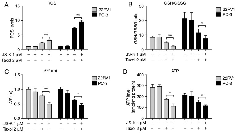
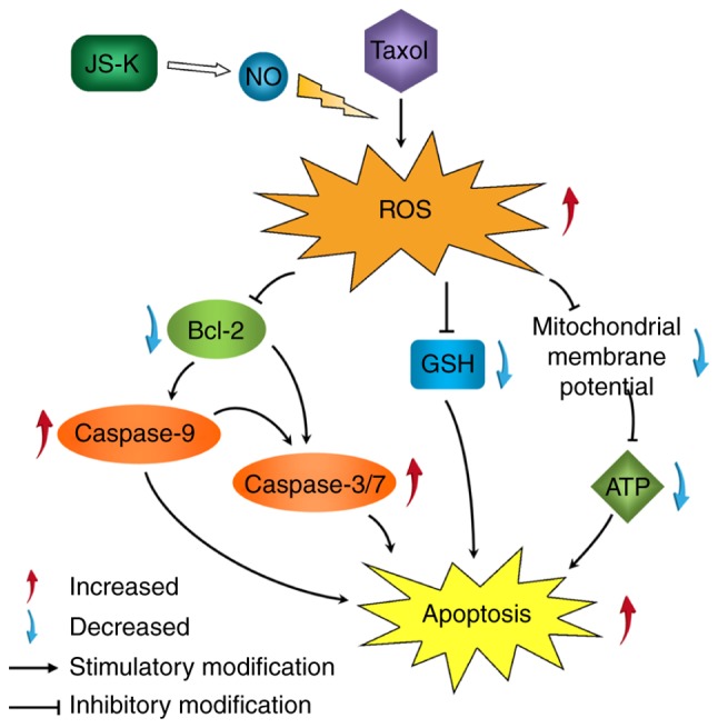
The aim of the present study was to investigate the influence of the nitric oxide donor prodrug JS-K (C13H16N6O8) on Taxol-induced apoptosis in prostate cancer cells, and to investigate a potential reactive oxygen species (ROS)-associated mechanism. The effect of JS-K on the anticancer activity of Taxol was assessed in prostate cancer cells; cell viability, colony formation, apoptosis, ROS generation and expression levels of apoptosis-associated proteins were investigated. The function of ROS accumulation in the combined effects of JS-K and Taxol was determined using the antioxidant N-acetylcysteine (NAC) and the pro-oxidant oxidized glutathione (GSSG). The results of the present study demonstrated that JS-K was able to increase Taxol-induced suppression of prostate cancer cell proliferation, apoptosis, ROS accumulation and upregulation of apoptosis-associated proteins. Furthermore, NAC reversed the effect of JS-K on Taxol-induced apoptosis and conversely, the pro-oxidant GSSG exacerbated the effect of JS-K on Taxol-induced apoptosis in prostate cancer cells. In conclusion, JS-K enhances the chemosensitivity of prostate cancer cells to Taxol, via the upregulation of intracellular ROS.

Keywords: JS-K; Taxol; chemosensitivity; prostate cancer cells; reactive oxygen species.


Figures

Figure 1.
Figure 1.
JS-K promotes Taxol-induced suppression of prostate cancer cell proliferation and apoptosis. (A) Cell viability following treatment with JS-K and Taxol was determined using a Cell Counting Kit-8 assay. (B) Representative images and (C) cumulative data from colony formation assay following treatment with JS-K and Taxol in 22RV1 and PC-3 cells. (D) Effects of JS-K on IC50 of Taxol. (E) Apoptosis induced by JS-K and Taxol was examined using FITC-annexin V/PI staining; representative dot plots (left panel) and cumulative data (right panel). The results are presented as the mean ± SD for three independent experiments. *P<0.05, **P<0.01. IC50, half-maximal inhibitory concentration; FITC, fluorescein isothiocyanate; PI, propidium iodide.
Figure 2.
Figure 2.
Synergistic effects of JS-K and Taxol on ROS production, GSH/GSSG ratio, mitochondrial membrane potential and ATP levels in prostate cancer cells. Prostate cancer cells were treated with JS-K and Taxol for 6 h and (A) ROS generation, (B) GSH/GSSG ratio, (C) mitochondrial membrane potential and (D) ATP levels were determined. The data are presented as the mean ± standard deviation for three independent experiments. *P<0.05, **P<0.01. GSH, reduced glutathione; GSSG, oxidized glutathione; ATP, adenosine triphosphate; ROS, reactive oxygen species; ΔΨ(m), mitochondrial membrane potential.
Figure 3.
Figure 3.
Effects of JS-K and Taxol on the apoptosis-associated signaling pathway. mRNA levels of Bcl-2, Bax and Bak in (A) 22RV1 and (B) PC-3. (C) Expression levels of apoptosis-associated proteins. (D) Activity of caspase-3/7 and (E) activity of caspase-9. The data are presented as the mean ± standard deviation for three independent experiments. *P<0.05, **P<0.01. Bak, BRI1-associated receptor kinase 1; Bax, Bcl-2-associated X protein; Bcl2, B-cell lymphoma 2; ROS, reactive oxygen species; ATP, adenosine triphosphate; GSH, reduced glutathione; GSSG, oxidized glutathione.
Figure 4.
Figure 4.
Effects of NAC and GSSG on JS-K- and Taxol-induced apoptosis in prostate cancer cells. (A) cell viability was measured using a Cell Counting Kit-8 assay. (B) Accumulation of intracellular ROS in prostate cancer cells. (C) Caspase-3/7 activity of 22RV1 and PC-3 cells. (D) Apoptosis of prostate cancer cells treated by JS-K and Taxol with or without NAC (100 µM) and GSSH (5 µM) were analyzed using FITC-annexin V/PI staining. Cumulative results are presented. (E) Representative dot plots of apoptosis of prostate cancer cells. The data are presented as the mean ± standard deviation for three independent experiments. *P<0.05, **P<0.01. NAC, N-acetylcysteine; GSSG, oxidized glutathione; ROS, reactive oxygen species; FITC, fluorescein isothiocyanate; PI, propidium iodide; A, annexin V.
Figure 5.
Figure 5.
Proposed cellular pathway of JS-K on Taxol-induced cell apoptosis in prostate cancer cells. JS-K promoted Taxol-induced prostate cancer cell apoptosis via the activation of caspase-3/7 and caspase-9 and suppression of ATP and GSH/GSSG ratio. The production of ROS induced by Taxol increased, which eventually led to apoptosis. ATP, adenosine triphosphate; GSH, reduced glutathione; GSSG, oxidized glutathione; ROS, reactive oxygen species; Bcl-2, B-cell lymphoma 2; Bax, Bcl-2-associated X protein.

References

    1. Weaver BA. How Taxol/paclitaxel kills cancer cells. Mol Biol Cell. 2014;25:2677–2681. doi: 10.1091/mbc.e14-04-0916. - DOI - PMC - PubMed
    1. Rowinsky EK, Cazenave LA, Donehower RC. Taxol: A novel investigational antimicrotubule agent. J Natl Cancer Inst. 1990;82:1247–1259. doi: 10.1093/jnci/82.15.1247. - DOI - PubMed
    1. Danesi R, Figg WD, Reed E, Myers CE. Paclitaxel (taxol) inhibits protein isoprenylation and induces apoptosis in PC-3 human prostate cancer cells. Mol Pharmacol. 1995;47:1106–1111. - PubMed
    1. Obasaju C, Hudes GR. Paclitaxel and docetaxel in prostate cancer. Hematol Oncol Clin North Am. 2001;15:525–545. doi: 10.1016/S0889-8588(05)70230-6. - DOI - PubMed
    1. Kopczyńska E. Role of microRNAs in the resistance of prostate cancer to docetaxel and paclitaxel. Contemp Oncol (Pozn) 2015;19:423–427. - PMC - PubMed