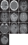
Ruptured intracranial dermoid cysts: a pictorial review
- PMID: 30655926
- PMCID: PMC6334092
- DOI: 10.5114/pjr.2018.80206
Ruptured intracranial dermoid cysts: a pictorial review
Abstract
Intracranial dermoid cysts are rare, benign, congenital, slow-growing cystic lesions. They are composed of mature squamous epithelium and can contain apocrine, eccrine, and sebaceous glands as well as other exodermal structures. Rupture of intracranial dermoid cysts is a relatively uncommon phenomenon but can cause more serious complications such as chemical meningitis, vasospasm, and cerebral infarction. Understanding of the appearance of both unruptured and ruptured dermoid cysts on computed tomography and MRI, especially awareness of existing low signal "blooming artefacts" on certain sequences, aids diagnosis and referral to the proper specialty for appropriate treatment.
Keywords: blooming artefacts; dermatoid cyst; magnetic resonance imaging; rupture.
Figures


References
-
- Osborn AG, Preece MT. Intracranial cysts: radiologic-pathologic correlation and imaging approach. Radiology. 2006;239:650–664. - PubMed
-
- Mehta MP, Chang SM, Guha A, et al. Principles and Practice of Neuro-Oncology: A Multidisciplinary Approach. Demos Medical; 2010.
-
- Runge VM, Smoker WRK, Valavanis A. Neuroradiology: The Essentials with MR and CT. Berlin: Thieme; 2014.
Publication types
LinkOut - more resources
Full Text Sources
