. 2018 Oct 26;3(1):49-58.
doi: 10.1002/rth2.12158. eCollection 2019 Jan.

Circulating endothelial cells as biomarker for cardiovascular diseases

Affiliations

Circulating endothelial cells as biomarker for cardiovascular diseases

Maura Farinacci et al. Res Pract Thromb Haemost. .

Abstract

Background: Endothelial dysfunction is involved in several cardiovascular diseases. Elevated levels of circulating endothelial cells (CECs) and low levels of endothelial progenitor cells (EPCs) have been described in different cardiovascular conditions, suggesting their potential use as diagnostic biomarkers for endothelial dysfunction. Compared to typical peripheral blood leukocyte subsets, CECs and EPCs occur at very low frequency. The reliable identification and characterization of CECs and EPCs is a prerequisite for their clinical use, however, a validated method to this purpose is still missing but a key for rare cell events.

Objectives: To establish a validated flow cytometric procedure in order to quantify CECs and EPCs in human whole blood.

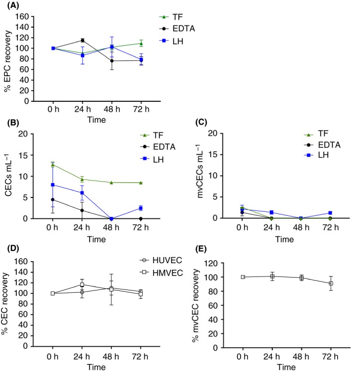
Methods: In the establishment phase, the assay sensitivity, robustness, and the sample storage conditions were optimized as prerequisite for clinical use. In a second phase, CECs and EPCs were analyzed in heart failure with preserved (HFpEF) and reduced (HFrEF) ejection fraction, in arterial hypertension (aHT), and in diabetic nephropathy (DN) in comparison to age-matched healthy controls.

Results: The quantification procedure for CECs and EPCs showed high sensitivity and reproducibility. CEC values resulted significantly increased in patients with DN and HFpEF in comparison to healthy controls. CEC quantification showed a diagnostic sensitivity of 90% and a sensitivity of 68.0%, 70.4%, and 66.7% for DN, HFpEF, and aHT, respectively.

Conclusion: A robust and precise assay to quantify CECs and EPCs in pre-clinical and clinical studies has been established. CEC counts resulted to be a good diagnostic biomarker for DN and HFpEF.

Keywords: biomarkers; cardiovascular diseases; endothelial cells; endothelial progenitor cells; flow cytometry.


Figures

Figure 1
Figure 1
Gating strategy for identification of CECs, EPCs, and for PMNC absolute count. The gating strategy for CEC or mvCECs (A, B) was as following: after gating out cell debris in the FSC/SSC dot‐plot and doublets in the FSC‐A/FSC‐H dot‐plot, DNA+ cells were identified as Syto16+, and leukocytes were excluded in the CD45/CD31 dot‐plot. Finally, CECs were identified as DNA+, CD45dim, CD31+CD146+ cells. mvCECs were then identified as CD36+ CECs. (A) Blood sample from a patient; (B) blood sample spiked with L‐HMVEC 1000 cells mL−1. For the detection of EPCs (C), cell debris were excluded in the FSC/SSC dot‐plot and doublets in the FSC‐A/FSC‐H dot‐plot, mononuclear cells were gated in the FSC/SSC plot and then subgated to identify the CD45dim CD34+ cells. From these cells, the CD45dimCD34+CD133+ EPCs were then identified. CD31 expression was analysed to further confirm EPCs identity. Absolute counts of CEC sand EPCs were determined multiplying the cell percentages relatively to the PMNCs by the absolute PMNC count obtained in separate tubes using Flow‐Count Fluorospheres (D). Here, blood samples were only stained with CD45; PMNC were identified by means of size and CD45 expression, the fluorospheres by means of size and fluorescence of PECy7, to exclude the PECy7‐blood cells. CECs, circulating endothelial cells; EPCs, endothelial progenitor cells
Figure 2
Figure 2
Performance of CEC detection and identification of microvascular cells. The graph in (A) shows the recovery rate of HUVEC and L‐HMVEC spiked in a healthy whole blood sample at 100, 1000 and 10 000 cells mL−1. The graph in (B) shows the percentage of CD36 positive cells detected on HMVEC and HUVEC at increasing spiking concentrations as in (A). The results are expressed as mean ± SD of duplicate quantification. CECs, circulating endothelial cells
Figure 3
Figure 3
Stability of EPCs (A), CECs (B, D), and mvCECs (C, E) in blood samples collected with different anticoagulants. Analyses were performed on fresh blood samples, 0 h, and after 24 h, 48 h and 72 h of storage at 4°C. EPC (A) and CEC (B, C) were quantified on whole blood samples collected from heathy donors in Transfix, EDTA or Lithium Heparin. Recovery of CECs and mvCECs (D, E) in Transfix tubes was assessed on whole blood samples spiked with HUVEC or L‐HMVEC at 100 cells mL−1 and expressed in percentage of time=0 h. Results are expressed as mean ± SD of two or three independent experiments. TF, transfix; LH, Lithium Heparin; CECs, circulating endothelial cells; EPCs, endothelial progenitor cells
Figure 4
Figure 4
CECs, EPCs and mvCECs in blood samples from patients with DN, HFpEF, HFrEF, aHT, and healthy individuals. Graphs of CECs (A), EPCs (B), and mvCECs (C) show median and interquartile range (grey bars). Statistical analysis was performed by ANOVA with Dunnett post‐test to compare each patient group to the healthy one. *P < 0.05. CECs, circulating endothelial cells; EPCs, endothelial progenitor cells; DN, diabetic nephropathy; HFpEF, heart failure with preserved ejection fraction; HFrEF, heart failure with reduced ejection fraction; aHT, arterial hypertension
Figure 5
Figure 5
Diagnostic sensitivity and specificity. Diagnostic sensitivity (A) and specificity (B) of CEC counts calculated for DN, HFpEF, HFrEF and aHT considering a cutoff at 10.5 cells mL−1. The table in (C) shows the levels of sensitivity and specificity obtained with cut‐off values higher or lower than 10.5. CEC, circulating endothelial cell; DN, diabetic nephropathy; HFpEF, heart failure with preserved ejection fraction; HFrEF, heart failure with reduced ejection fraction; aHT, arterial hypertension

References

    1. Lozano R, Naghavi M, Foreman K, et al. Global and regional mortality from 235 causes of death for 20 age groups in 1990 and 2010: a systematic analysis for the Global Burden of Disease Study 2010. Lancet. 2012;380:2095–128. - PMC - PubMed
    1. Endemann DH, Schiffrin EL. Endothelial dysfunction. J Am Soc Nephrol. 2004;15:1983–92. - PubMed
    1. Peters K, Unger RE, Brunner J, Kirkpatrick CJ. Molecular basis of endothelial dysfunction in sepsis. Cardiovasc Res. 2003;60:49–57. - PubMed
    1. Thambyrajah J, Landray MJ, McGlynn FJ, Jones HJ, Wheeler DC, Townend JN. Abnormalities of endothelial function in patients with predialysis renal failure. Heart. 2000;83:205–9. - PMC - PubMed
    1. Landray MJ, Wheeler DC, Lip GY, et al. Inflammation, endothelial dysfunction, and platelet activation in patients with chronic kidney disease: the chronic renal impairment in Birmingham (CRIB) study. Am J Kidney Dis. 2004;43:244–53. - PubMed