

Electroconvulsive treatment prevents chronic restraint stress-induced atrophy of the hippocampal formation-A stereological study
- PMID: 30656862
- PMCID: PMC6379514
- DOI: 10.1002/brb3.1195
Electroconvulsive treatment prevents chronic restraint stress-induced atrophy of the hippocampal formation-A stereological study
Abstract
Introduction: Electroconvulsive therapy (ECT) is one of the most efficient treatments of major depressive disorder (MDD), although the underlying neurobiology remains poorly understood. There is evidence that ECT and MDD exert opposing effects on the hippocampal formation with respect to volume and number of neurons. However, there has been a paucity of quantitative data in experimental models of ECT and MDD.
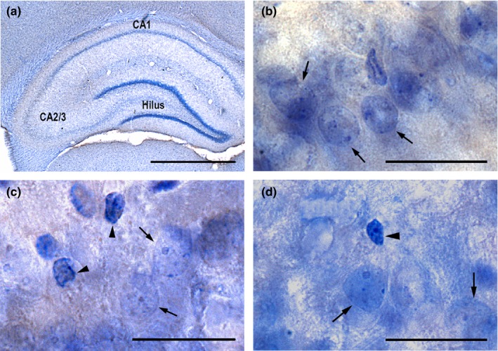
Methods: Using design-based stereology, we have measured the effects of a stress-induced depression model (chronic restraint stress, CRS) and ECS on the morphology of the hippocampus by estimating the volume and total number of neurons in the hilus, CA1, and CA2/3, as well as in the entire hippocampus.
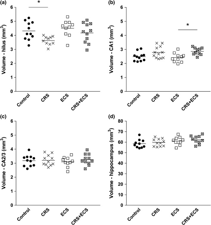
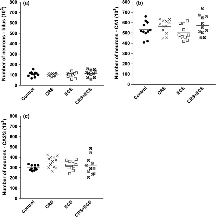
Results: We find that CRS induces a significant decrease in volume exclusively of the hilus and that ECS (CRS + ECS) blocks this reduction. Furthermore, ECS alone does not change the volume or total number of neurons in the entire hippocampus or any hippocampal subdivision in our rat model.
Keywords: cell numbers; chronic restraint stress; electroconvulsive stimulation; hippocampal volumes.
© 2019 The Authors. Brain and Behavior published by Wiley Periodicals, Inc.
Figures
References
-
- Boldrini, M. , Santiago, A. N. , Hen, R. , Dwork, A. J. , Rosoklija, G. B. , Tamir, H. , … Mann, J. (2013). Hippocampal granule neuron number and dentate gyrus volume in antidepressant‐treated and untreated major depression. Neuropsychopharmacology, 38, 1068–1077. 10.1038/npp.2013.5 - DOI - PMC - PubMed
-
- Calfa, G. , Kademian, S. , Ceschin, D. , Vega, G. , Rabinovich, G. A. , & Volosin, M. (2003). Characterization and functional significance of glucocorticoid receptors in patients with major depression: Modulation by antidepressant treatment. Psychoneuroendocrinology, 28, 687–701. 10.1016/S0306-4530(02)00051-3 - DOI - PubMed
Publication types
MeSH terms
LinkOut - more resources
Full Text Sources
Miscellaneous
