. 2019 Jan 18;18(1):10.
doi: 10.1186/s12934-019-1058-4.

Engineering the flagellar type III secretion system: improving capacity for secretion of recombinant protein

Affiliations

Engineering the flagellar type III secretion system: improving capacity for secretion of recombinant protein

Charlotte A Green et al. Microb Cell Fact. .

Abstract

Background: Many valuable biopharmaceutical and biotechnological proteins have been produced in Escherichia coli, however these proteins are almost exclusively localised in the cytoplasm or periplasm. This presents challenges for purification, i.e. the removal of contaminating cellular constituents. One solution is secretion directly into the surrounding media, which we achieved via the 'hijack' of the flagellar type III secretion system (FT3SS). Ordinarily flagellar subunits are exported through the centre of the growing flagellum, before assembly at the tip. However, we exploit the fact that in the absence of certain flagellar components (e.g. cap proteins), monomeric flagellar proteins are secreted into the supernatant.

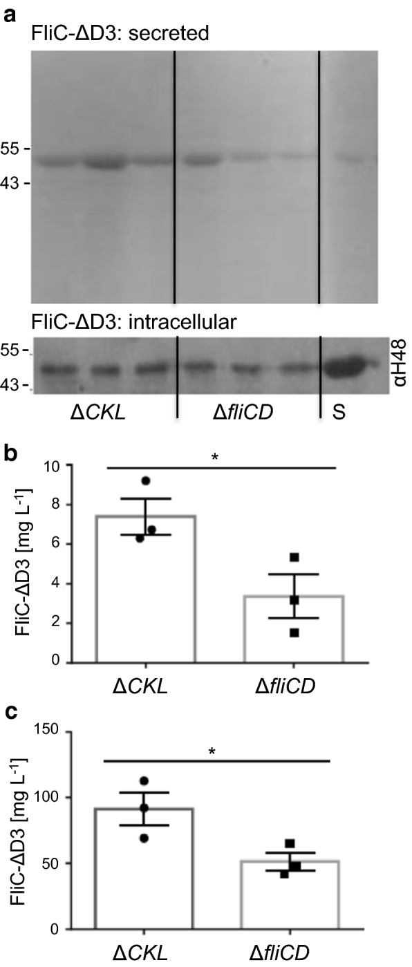
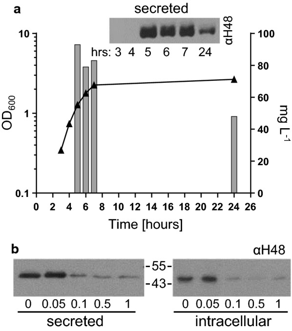
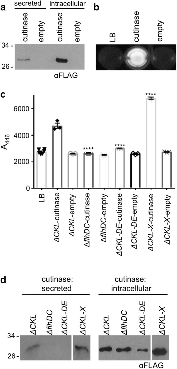
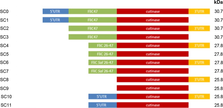
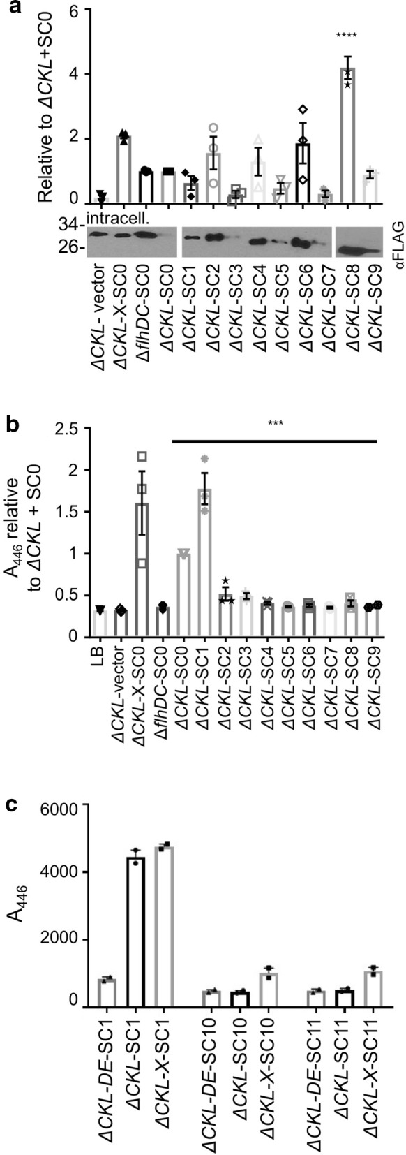
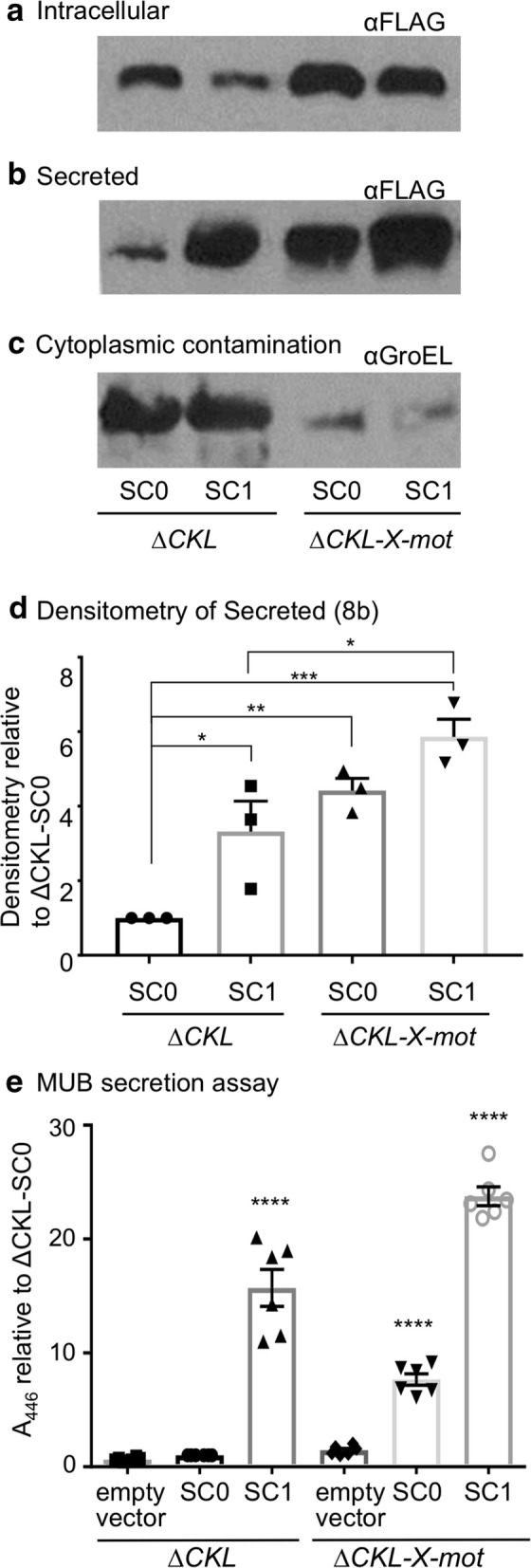
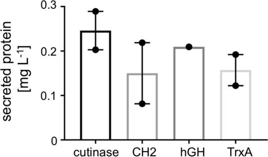
Results: We report the creation and iterative improvement of an E. coli strain, by means of a modified FT3SS and a modular plasmid system, for secretion of exemplar proteins. We show that removal of the flagellin and HAP proteins (FliC and FlgKL) resulted in an optimal prototype. We next developed a high-throughput enzymatic secretion assay based on cutinase. This indicated that removal of the flagellar motor proteins, motAB (to reduce metabolic burden) and protein degradation machinery, clpX (to boost FT3SS levels intracellularly), result in high capacity secretion. We also show that a secretion construct comprising the 5'UTR and first 47 amino acidsof FliC from E. coli (but no 3'UTR) achieved the highest levels of secretion. Upon combination, we show a 24-fold improvement in secretion of a heterologous (cutinase) enzyme over the original strain. This improved strain could export a range of pharmaceutically relevant heterologous proteins [hGH, TrxA, ScFv (CH2)], achieving secreted yields of up to 0.29 mg L-1, in low cell density culture.

Conclusions: We have engineered an E. coli which secretes a range of recombinant proteins, through the FT3SS, to the extracellular media. With further developments, including cell culture process strategies, we envision further improvement to the secreted titre of recombinant protein, with the potential application for protein production for biotechnological purposes.

Keywords: Biotechnology; Flagellar; Recombinant protein secretion; Secretion assay; Strain engineering; Synthetic biology.


Figures

Fig. 1
Fig. 1
Comparison of secretion capacity in the HAP-less and cap-less strains. E. coli MC1000 ΔfliC ΔflgKL (ΔCKL) and ΔfliCD containing the plasmid pTrc-FliC-ΔD3 were grown in LB supplemented with 0.05 mM IPTG, and harvested at OD600 1.5. Samples which represented either 25 µL or 400 µL culture media were loaded for SDS-PAGE, for intracellular and secreted protein respectively. a Representative images showing the secreted fractions following Coomassie staining, and the intracellular fractions following immunoblotting using an anti-flagellin (H48) antibody and a HRP secondary. A FliC-ΔD3 protein standard (S) was also included, to allow quantification of protein concentration by densitometry (b, c). Quantifications for biological triplicates, ± SE and individual data points shown, *p < 0.05
Fig. 2
Fig. 2
Optimisation of cell culture induction and harvesting procedure for the FliC-ΔD3 protein secretion assay. The ‘prototype’ E. coli MC1000 ΔfliC ΔflgKL (ΔCKL) containing the plasmid pTrc-FliC-ΔD3 was grown in 100 mL flask cultures in LB and either a supplemented with 0.05 mM IPTG and harvested every hour or b supplemented with 0, 0.05, 0.1, 0.5 or 1 mM IPTG and harvested at OD600 1.0. FliC-ΔD3 was detected by immunoblot as described in Fig. 1a. Samples which represented either 25 µL or 300 µL culture media were loaded for SDS-PAGE for intracellular and secreted protein respectively. Densitometry analysis allowed quantification of secreted protein, throughout the growth curve. This was repeated to ensure that this corroborated with the general trend
Fig. 3
Fig. 3
Protein secretion through the truncated FT3SS can be both controlled and improved. E. coli MC1000 ΔfliC ΔflgKL (ΔCKL or ‘prototype’), ΔflhDC, ΔfliC ΔflgKL ΔflgDE (ΔCKL-DE) or ΔfliC ΔflgKL ΔclpX (ΔCKL-X or ‘Mark II strain’) containing pTrc-FliC-ΔD3 was grown with 0.05 mM IPTG (or 1 mM to allow overexpression in ΔflhDC) and harvested at OD600 1.0. Secreted and intracellular cell fractions were loaded for SDS-PAGE in the quantities described in Fig. 2. A FliC-ΔD3 protein standard was included to allow quantification. Samples underwent a Coomassie staining or immunoblot analysis of cells and supernatant using b anti-flagellin (αH48) or c anti(α)-GroEL primary antibodies and a HRP-conjugated secondary
Fig. 4
Fig. 4
Development of a high throughput fluorescence assay to measure protein secretion through the truncated FT3SS. The ‘prototype’ E. coli MC1000 ΔfliC ΔflgKL (ΔCKL) containing either pJex-fliC47-cutinase or pJex-fliC47-empty were grown as described in Fig. 3 and prepared for a immunoblot with the anti-FLAG-HRP (αFLAG) antibody (where samples representing either 15 µL or 300 µL culture media for intracellular and secreted protein respectively were loaded onto the SDS-PAGE) (b) or florescence assay: 40 µL supernatant was added to 160 µL MUB substrate in a 96 well plate. Following incubation for 30 min at 30 °C, samples were visualised under UV light. c, d E. coli strains ΔfliC ΔflgKL (ΔCKL or ‘prototype’), ΔfliC ΔflgKLclpX (ΔCKL-X or ‘Mark II strain’), ∆flhDC and ΔfliC ΔflgKLflgDE (ΔCKL-DE) harbouring the cutinase expressing or empty vector plasmid were grown and c prepared for MUB secretion assay as described above; however following incubation, fluorescence was measured in a plate reader (excitation 302 nm, emission 446 nm). Results from one biological replicate, with three technical repeats. ± SE and individual data points shown. Two-way ANOVA (variables: strain and plasmid) and Tukey’s multiple comparison test: ****p < 0.001. d Representative immunoblot of secreted and intracellular fractions prepared from the same cell cultures
Fig. 5
Fig. 5
Screening for strains which are high capacity secretors of recombinant cutinase, and additional substrates. a The ‘prototype’ E. coli MC1000 ΔfliC ΔflgKL (ΔCKL) strain with additional combinations of: ΔmotAB (mot) ΔflgMN (MN) ΔfliDST (DST) or ΔclpX (X), which contained pJex-fliC47-cutinase were grown with 0.05 mM IPTG and harvested at OD600 1.0. Secreted fractions (Sn) were analysed by the MUB florescence assay as described in Fig. 4c. Results from three biological replicates were normalised to ΔCKL, ± SE shown, one-way ANOVA: p ≤ 0.001. Tukey’s multiple comparison test to ΔCKL: *p ≤ 0.05, **p ≤ 0.01. Representative immunoblot of the intracellular fraction from 15 µL cell culture shown below. b E. coli ΔCKL (‘prototype’), ΔCKL-X-mot (Mark III strain) and ΔCKL-mot-DST expressing pJex-fliC47-CH2 (+, upper), pTrc-FliC-ΔD3 (+, lower), or empty vector (−) were grown (as above) and prepared for immunoblot using either an anti-FLAG-HRP (αFLAG) antibody (upper) or an anti-flagellin (αH48) antibody and a HRP conjugated secondary (lower). The equivalent of 15 µL cell culture was loaded for intracellular fractions, and either 300 or 60 µL for the secreted fractions (for CH2 and FliC-ΔD3 respectively), along with the relevant protein standard to allow quantification. c Whole cell fractions underwent immunoblot with an anti-FlhA antibody (αFlhA), results from densitometry are presented relative to ΔCKL. Five biological repeats, ± SE and individual data points shown. One-way ANOVA: p < 0.005 and Tukey’s multiple comparison test: *p < 0.05, **p < 0.01
Fig. 6
Fig. 6
Schematic of secretion signal variants. The prototype secretion construct (SC0), and variations (SC1–SC11. Note that SC1 contributes to the ‘Mark IV’ improvement) are depicted along with the predicted size of the protein product (kDa). All secretion constructs harbour cutinase, along with combinations of the 3′UTR, 5′UTR, the 1–47 residue FliC secretion signal, or the truncated secretion signal (residues 26–47) with the native E. coli or S. typhimurium codon usage. Construction of these plasmids is outlined in Additional file 1: Fig. S5 and Table S2
Fig. 7
Fig. 7
Comparison of expression and secretion in secretion signal variants. a, b The ‘prototype’ E. coli ΔfliC ΔflgKL (ΔCKL) harbouring either pJex-fliC47-empty or the secretion signal variants SC0–SC9 (see Fig. 6), along with ΔflhDC and ΔfliC ΔflgKL ΔclpX (ΔCKL-X or ‘Mark II strain’) expressing SC0 were grown and harvested as in Fig. 3. a Intracellular fractions were prepared for immunoblotting (plus densitometry analysis: representative image shown), and b secreted fractions for MUB secretion assay, as described in Fig. 4a and c respectively. Three biological repeats, normalised to ΔCKL-SC0, ± SE and individual data points shown. One-way ANOVA (for SC0–SC9 expressing strains only) p < 0.001 and Tukey’s multiple comparison test (to ΔCKL-SC0): ****p < 0.001, ***p < 0.005. c The absence of the 47 residue FliC signal, with the presence of the fliC 5′UTR was investigated in ΔCKL, ΔCKL-X and ΔflhDC compared to SC1. Procedure as described for Fig. 7b. Two biological replicates, ± SE and individual data points shown
Fig. 8
Fig. 8
Cutinase expression and secretion: comparing the prototype, and most improved, strains and secretion constructs. The E. coli ‘prototype’ ΔfliC ΔflgKL (ΔCKL) or ‘Mark III strain’ ΔfliC ΔflgKL ΔclpX ΔmotAB (ΔCKL-X-mot) expressing either the prototype (SC0), improved (SC1) modular secretion vector, or pJex–fliC47-empty (empty), were grown as described in Fig. 3. Note that ΔCKL-X-mot expressing SC1 is our ‘Mark IV strain’. Both intracellular and secreted fractions underwent immunoblot analysis using either, an anti-FLAG-HRP (αFLAG) antibody to detect a intracellular and b secreted cutinase, or c anti-GroEL and a HRP secondary antibody to detect cytoplasmic protein contamination. Samples representing 15 μL and 300 μL cell culture were loaded for intracellular and supernatant samples, respectively. d Densitometry analysis was carried out on αFLAG probed, secreted fractions (Fig. 8b: representative image) of three biological replicates, normalised to ΔCKL-SC0, ± SE and individual data points shown. One-way ANOVA, p < 0.005 and Tukey’s multiple comparison test (to ΔCKL-SC0): ***p < 0.005, **p < 0.01, *p < 0.05. e Supernatant was also prepared for MUB protein secretion assay as described in Fig. 4c. Six biological replicates, normalised to ΔCKL-SC0, ± SE and individual data points shown. Two-way ANOVA, p < 0.001 and Tukey’s multiple comparison test (to ΔCKL-SC0): ****p < 0.001
Fig. 9
Fig. 9
Secreted titres of a range of substrates through the optimised Mark IV secretion strain. E. coli ΔfliC ΔflgKL ΔclpX ΔmotAB expressing plasmid based SC1 (Mark IV strains), with either cutinase, CH2, hGH or TrxA cargo, were grown as outlined in Fig. 3 and prepared, along with a relevant protein standard (exception: cutinase, where a hGH standard was utilised), for immunoblot detection with anti-FLAG-HRP (αFLAG). The concentration of secreted protein was then quantified by densitometry analysis. Two biological replicates (with the exception of hGH), ± SE and individual data points shown

Similar articles

Cited by

References

    1. Ferrer-Miralles N, Villaverde A. Bacterial cell factories for recombinant protein production; expanding the catalogue. Microb Cell Fact. 2013;12:113. doi: 10.1186/1475-2859-12-113. - DOI - PMC - PubMed
    1. Baeshen MN, Al-Hejin AM, Bora RS, Ahmed MMM, Ramadan HAI, Saini KS, et al. Production of Biopharmaceuticals in E. coli: current scenario and future perspectives. J Microbiol Biotechnol. 2015;25:953–962. doi: 10.4014/jmb.1412.12079. - DOI - PubMed
    1. Hellwig S, Drossard J, Twyman RM, Fischer R. Plant cell cultures for the production of recombinant proteins. Nat Biotechnol. 2004;22:1415–1422. doi: 10.1038/nbt1027. - DOI - PubMed
    1. Walsh G. Biopharmaceutical benchmarks 2014. Nat Biotechnol. 2014;32:992–1000. doi: 10.1038/nbt.3040. - DOI - PubMed
    1. Freudl R. Signal peptides for recombinant protein secretion in bacterial expression systems. Microb Cell Fact. 2018;17:52. doi: 10.1186/s12934-018-0901-3. - DOI - PMC - PubMed

MeSH terms