. 2019 Mar;110(3):962-972.
doi: 10.1111/cas.13946. Epub 2019 Feb 8.

REV7 confers radioresistance of esophagus squamous cell carcinoma by recruiting PRDX2

Affiliations

REV7 confers radioresistance of esophagus squamous cell carcinoma by recruiting PRDX2

Cheng Gu et al. Cancer Sci. 2019 Mar.

Abstract

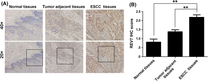
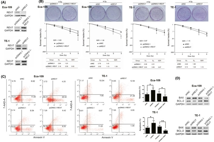
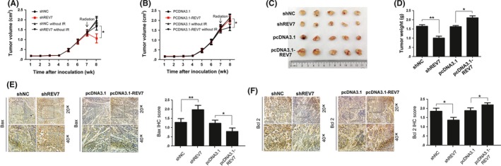
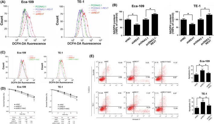
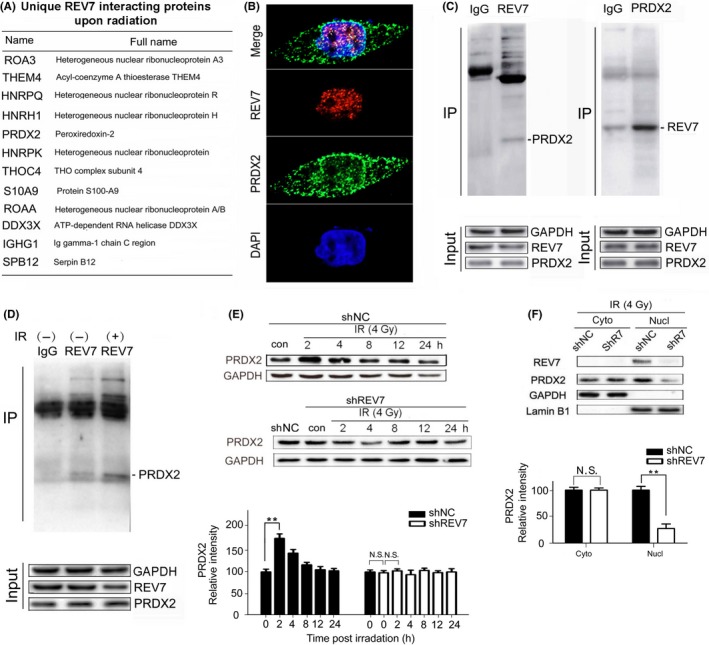
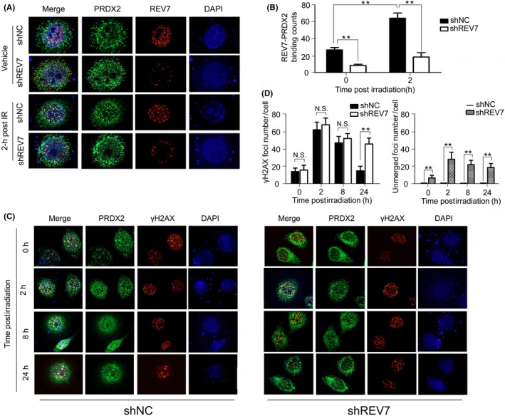
Radiotherapy has been widely used for the clinical management of esophageal squamous cell carcinoma. However, radioresistance remains a serious concern that prevents the efficacy of esophageal squamous cell carcinoma (ESCC) radiotherapy. REV7, the structural subunit of eukaryotic DNA polymerase ζ, has multiple functions in bypassing DNA damage and modulating mitotic arrest in human cell lines. However, the expression and molecular function of REV7 in ESCC progression remains unclear. In this study, we first examined the expression of REV7 in clinical ESCC samples, and we found higher expression of REV7 in ESCC tissues compared to matched adjacent or normal tissues. Knockdown of REV7 resulted in decreased colony formation and increased apoptosis in irradiated Eca-109 and TE-1 cells coupled with decreased tumor weight in a xenograft nude mouse model postirradiation. Conversely, overexpression of REV7 resulted in radioresistance in vitro and in vivo. Moreover, silencing of REV7 induced increased reactive oxygen species levels postirradiation. Proteomic analysis of REV7-interacting proteins revealed that REV7 interacted with peroxiredoxin 2 (PRDX2), a well-known antioxidant protein. Existence of REV7-PRDX2 complex and its augmentation postirradiation were further validated by immunoprecipitation and immunofluorescence assays. REV7 knockdown significantly disrupted the presence of nuclear PRDX2 postirradiation, which resulted in oxidative stress. REV7-PRDX2 complex also assembled onto DNA double-strand breaks, whereas REV7 knockdown evidently increased double-strand breaks that were unmerged by PRDX2. Taken together, the present study sheds light on REV7-modulated radiosensitivity through interacting with PRDX2, which provides a novel target for ESCC radiotherapy.

Keywords: DNA double-strand breaks; PRDX2; REV7; radioresistance; reactive oxygen species.


Conflict of interest statement

The authors have declared no conflicts of interest to this work.

Figures

Figure 1
Figure 1
Higher expression of REV7 in esophageal squamous cell carcinoma (ESCC) samples. A, Representative immunohistochemistry (IHC) staining of REV7 expression in ESCC tissue, tumor‐adjacent tissue and normal esophageal tissue specimens (magnification 20× or 40×). B, Bar plot representing the IHC staining score of REV7 in ESCC tissues (n = 102), tumor‐adjacent tissues (n = 52) and normal esophageal tissues (n = 21). **< .01
Figure 2
Figure 2
REV7 confers radioresistance on esophageal squamous cell carcinoma cells. A, Western blotting analysis of REV7 expression in Eca‐109 and TE‐1 cells transfected with shREV7, shNC, pcDNA3.1 or pcDNA3.1‐REV7. GAPDH served as an internal control. B, Colony formation of REV7‐overexpressing or REV7‐knockdown Eca‐109 and TE‐1 cells. Clonogenic survival curves were generated for Eca‐109 and TE‐1 cells that were stably transfected with the indicated vectors and were then exposed to 2, 4, 6 or 8 Gy X‐ray irradiation. The D0, Dq and sensitization enhancement ratio (SER) value of the corresponding groups is shown. The survival curve was derived from a multi‐target single‐hit model: SF = 1‐1‐exp(‐D/D0)n. D0 was defined as the dose that gave an average of 1 hit per target. Dq represents the repair of nonlethal injury, a higher Dq value means a higher dose is required to cause the death of cells. The SER was measured according to the multi‐target single‐hit model. C, Apoptosis rates were measured using Annexin‐V/7‐AAD staining in Eca‐109 and TE‐1 cells treated with the indicated vectors at 48 h after 4 Gy X‐ray irradiation. The data are shown as the mean ± SEM of 3 independent experiments. *P < .05, **P < .01. D, Western blotting analysis of Bcl‐2 and Bax expression for indicated group
Figure 3
Figure 3
REV7 modulates the radioresistance of esophageal squamous cell carcinoma (ESCC) tumor xenografts. A‐B, Growth curve of Eca109 in subcutaneous tumor xenografts. C‐D, Representative images of the volume and weight of tumors originating from ESCC tumor xenografts in nude mice at 8 wk (1 wk postirradiation). The tumor weights are presented as the mean ± SEM of 3 independent experiments. **< .01, *< .05. E‐F, Representative immunohistochemistry (IHC) staining of Bax and Bcl2 in the indicated tumor xenografts. Scores or counts of IHC were evaluated by 2 independent observers in a blinded manner and are presented as the mean ± SEM of 3 independent experiments. **< .01, *< .05
Figure 4
Figure 4
REV7 maintains the redox balance in irradiated esophageal squamous cell carcinoma cells. A, Representative curves of the reactive oxygen species (ROS) level, as indicated by DCFH fluorescence probe staining, were obtained using flow cytometric analysis of Eca109/TE‐1 cells 2 h after 4 Gy of X‐ray irradiation. B, Corresponding relative NADPH content was assayed by NADPH kit, presented as the mean ± SEM of 3 independent experiments. **< .01,*< .05. C, Representative curves of ROS level in REV7 KD and REV7 KD + NAC Eca109 cells. D, Colony formation of REV7 KD and REV7 KD + NAC Eca109 cells. E, Apoptosis rates were measured using Annexin‐V/7‐AAD staining in REV7 KD and REV7 KD + NAC Eca‐109 cells at 48 h after 4 Gy X‐ray irradiation. The data are shown as the mean ± SEM of 3 independent experiments. *P < .05
Figure 5
Figure 5
REV7 interacts with PRDX2 in the nucleus of esophageal squamous cell carcinoma cells. A, List of unique proteins that increased in percentage based on proteomic analysis at 2 h after 4 Gy X‐ray irradiation. B, Immunofluorescent staining of REV7 and PRDX2 in Eca‐109 cells (coexistence of REV7 and PRDX2 are indicated by the arrows). C, Confirmation of PRDX2/REV7 protein co‐immunoprecipitation by beads containing REV7/PRDX2 antibody using whole cell lysate from Eca‐109 cells, as revealed by western blot analysis. D, Immunoprecipitation assay by beads containing REV7 antibody in whole cell lysate from Eca‐109 cell with or without irradiation (2 h after 4 Gy X‐ray), as revealed by western blot analysis. E, Western blotting analysis of PRDX2 expression of shNC/shREV7 group postirradiation. Relative intensity of PRDX2 normalized to GAPDH is presented as the mean ± SEM of 3 independent experiments. **P < .01, N.S. nonsignificant. F, Cytosolic and nuclear fractions isolated from shNC/shREV7 Eca109 cells were assayed by western blotting for PRDX2. Relative intensity of PRDX2 normalized to GAPDH is presented as the mean ± SEM of 3 independent experiments. **P < .01, N.S. nonsignificant
Figure 6
Figure 6
REV7‐PRDX2 complex is recruited onto double‐strand breaks (DSB) postirradiation. A‐B, Immunofluorescent staining of REV7 and PRDX2 in shNC/shREV7 Eca‐109 cells with or without irradiation (2 h after 4 Gy X‐ray). Bindings of REV7‐PRDX2 complex are observed by 2 independent observers in a blinded manner and are regarded as positive only if both observers counted independently and are presented as the mean ± SEM of 3 independent experiments. **P < .01. C‐D, Immunofluorescent staining of PRDX2 and γH2AX in shNC/shREV7 Eca‐109 cells with radiation (0, 2, 8 and 24 h after 4 Gy X‐ray). γH2AX foci merged or unmerged with nuclear PRDX2 were observed and calculated as indicated above. N.S., nonsignificant. ** P < .01, N.S. nonsignificant

Similar articles

Cited by

References

    1. Siegel RL, Miller KD, Jemal A. Cancer statistics, 2017. CA Cancer J Clin. 2017;67:7‐30. - PubMed
    1. Chung CS, Lee YC, Wu MS. Prevention strategies for esophageal cancer: perspectives of the East versus West. Best Pract Res Clin Gastroenterol. 2015;29:869‐883. - PubMed
    1. Cunningham D, Allum WH, Stenning SP, et al. Perioperative chemotherapy versus surgery alone for resectable gastroesophageal cancer. N Engl J Med. 2006;355:11‐20. - PubMed
    1. van Hagen P, Hulshof MC, Van Lanschot JJB, et al. Preoperative chemo‐radiotherapy for esophageal or junctional cancer. N Engl J Med. 2012;366:2074‐2084. - PubMed
    1. Suh YG, Lee IJ, Koom WS, et al. High‐dose versus standard‐dose radiotherapy with concurrent chemotherapy in stages II–III esophageal cancer. Jpn J Clin Oncol. 2014;44:534‐540. - PubMed

MeSH terms