. 2019 Mar;105(3):519-530.
doi: 10.1002/JLB.1AB0417-160RR. Epub 2019 Jan 18.

The mannose 6-phosphate/insulin-like growth factor 2 receptor mediates plasminogen-induced efferocytosis

Affiliations

The mannose 6-phosphate/insulin-like growth factor 2 receptor mediates plasminogen-induced efferocytosis

Anna Ohradanova-Repic et al. J Leukoc Biol. 2019 Mar.

Abstract

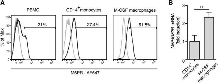
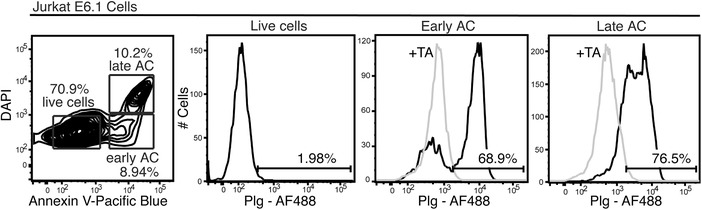
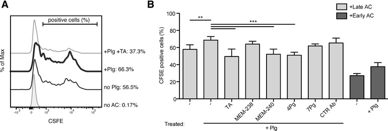
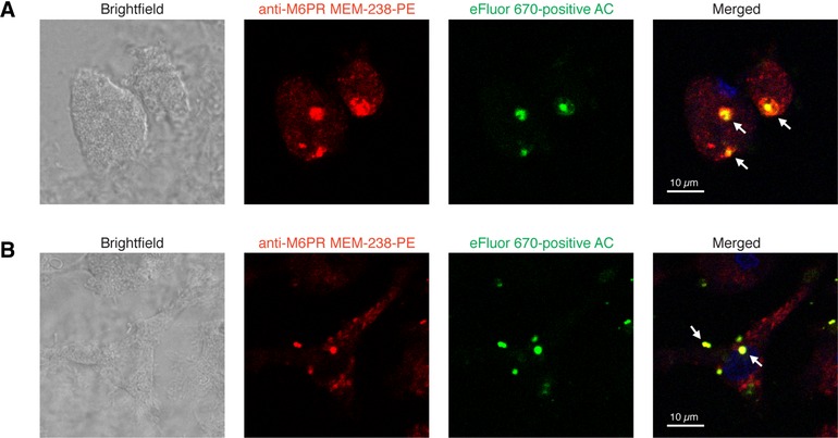
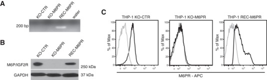
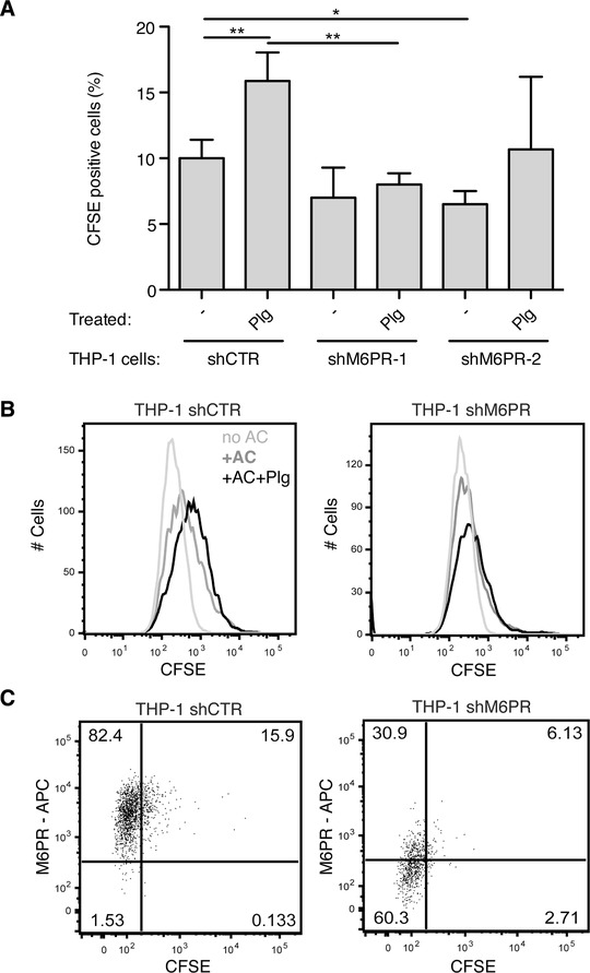
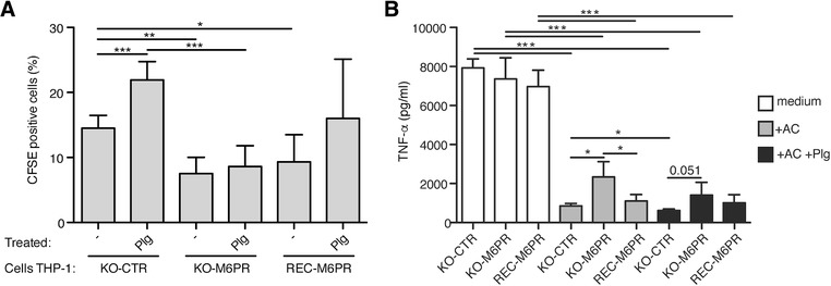
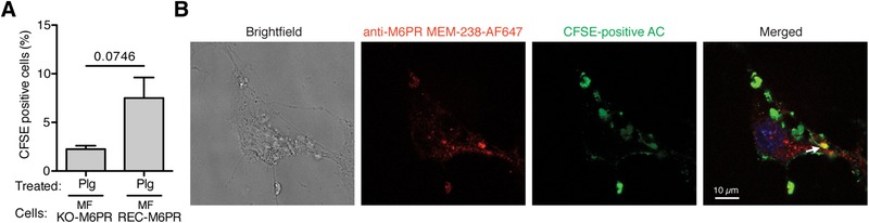
The plasminogen system is harnessed in a wide variety of physiological processes, such as fibrinolysis, cell migration, or efferocytosis; and accordingly, it is essential upon inflammation, tissue remodeling, wound healing, and for homeostatic maintenance in general. Previously, we identified a plasminogen receptor in the mannose 6-phosphate/insulin-like growth factor 2 receptor (M6P/IGF2R, CD222). Here, we demonstrate by means of genetic knockdown, knockout, and rescue approaches combined with functional studies that M6P/IGF2R is up-regulated on the surface of macrophages, recognizes plasminogen exposed on the surface of apoptotic cells, and mediates plasminogen-induced efferocytosis. The level of uptake of plasminogen-coated apoptotic cells inversely correlates with the TNF-α production by phagocytes indicating tissue clearance without inflammation by this mechanism. Our results reveal an up-to-now undetermined function of M6P/IGF2R in clearance of apoptotic cells, which is crucial for tissue homeostasis.

Keywords: M6P/IGF2R; efferocytosis; macrophages; plasminogen; tissue homeostasis.


Figures

Figure 1
Figure 1
M6P/IGF2R expression increases during monocyte differentiation to macrophages. (A) Cell‐surface expression of M6P/IGF2R on isolated PBMC and MACS‐enriched monocytes from healthy donors was evaluated with mAb MEM‐238‐AF647 and flow cytometry. In parallel, MOPC‐21‐AF647 was used as an isotype control mAb, displayed by the cut‐off gates. The same analysis was performed with macrophages differentiated from MACS‐sorted monocytes during a 7‐day culture with recombinant human M‐CSF (50 ng/ml) followed by 2 days resting in macrophage serum free medium. (B) Primary human monocytes and monocyte‐derived macrophages from (A) were lysed and RNA was extracted. cDNA was synthesized from total RNA and gene expression was measured by real‐time PCR as described in the Material and Methods section with TaqMan primer sets for human M6P/IGF2R and YWHAZ as endogenous control. The M6P/IGF2R mean expression values relative to that of monocytes ± SD from 3 donors is shown
Figure 2
Figure 2
Plg marks apoptotic cells. Jurkat T cells were stained on ice with Plg‐AF647, Annexin V‐Pacific blue and DAPI, and analyzed by flow cytometry to discriminate early (Annexin V+) and late (Annexin V+ / DAPI+) apoptotic cells (AC) from viable (Annexin V / DAPI) cells. Optionally, we co‐incubated the cells with Plg‐AF647 and TA (5 mmol/l)
Figure 3
Figure 3
Flow cytometry analysis of Plg‐mediated efferocytosis by human macrophages. (A) A representative flow cytometry histogram of the efferocytosis analysis. Jurkat T cells were fluorescently labeled with CFSE and their apoptosis was induced by SSP treatment (200 ng/ml) for 16 h. Then, the apoptotic cells (AC) were pretreated for 30 min with or without Plg (100 nmol/l) and TA (5 mmol/l), washed, and added to monocyte‐derived macrophages (generated as in Fig. 1). Incubation was performed for 2 h at 37°C at the macrophage:apoptotic cell ratio of 1:5; without Plg (thin black line), with Plg (bold black line), with Plg and TA (thin grey line). (B) Flow cytometry was used to quantify percentages of macrophages that phagocytosed CFSE‐labeled apoptotic cells. The levels of efferocytosis are displayed as percentages of CFSE‐positive macrophages. Incubation was performed for 2 h at 37°C at the macrophage:apoptotic cell ratio of 1:5 in the presence of the following additives: Plg, 100 nmol/l, TA, 5 mmol/l, mAbs MEM‐238 and MEM‐240 to M6P/IGF2R, mAbs 4Pg and 7Pg to Plg, and control mAb AFP‐01, all 5 μg/ml. In some experiments, apoptosis of the Jurkat cells was induced by SSP treatment (200 ng/ml) for 9 h (in dark grey). Mean ± SD of at least 5 independent experiments is shown
Figure 4
Figure 4
Confocal microscopy analysis of Plg‐mediated efferocytosis by human macrophages. (A) After incubation with eFluor 670‐labeled apoptotic Jurkat T cells (green) for 2 h the macrophages were fixed, permeabilized and stained with PE‐conjugated M6P/IGF2R mAb MEM‐238 (red). Nuclei were stained with DAPI (blue). The slides were washed and analyzed by confocal microscopy. (B) Macrophages grown on microscope slides were preloaded for 30 min with mAb MEM‐238‐PE, washed and subjected to the efferocytosis assay with eFluor 670‐labeled apoptotic Jurkat cells for 2 h. Then the slides were washed and analyzed by confocal microscopy; the scales represent 10 μm; arrows point to the region of colocalization of M6P/IGF2R and apoptotic cells (AC)
Figure 5
Figure 5
Generation of M6P/IGF2R‐knock‐out and ‐reconstituted THP‐1 cells. Knockout of M6P/IGF2R in THP‐1 cells was performed via targeting the M6P/IGF2R gene in the Exon 22 on chromosome 6 by means of the Zinc finger nuclease (ZFN) technology. The expression of M6P/IGF2R was reconstituted in the knockout cells through retroviral transduction of the DNA fragment encoding human M6P/IGF2R. The expression of M6P/IGF2R was verified at both the gene and the protein level. (A) Genomic DNA purified from THP‐1 control cells (KO‐CTR), M6P/IGF2R‐knock‐out cells (KO‐M6PR), and KO‐M6PR cells reconstituted with M6P/IGF2R (REC‐M6PR) was used as a template for the PCR amplification with exon‐crossing primer sets for human M6P/IGF2R recognizing the recombinant M6P/IGF2R gene. (B) The total M6P/IGF2R contents in control cells (KO‐CTR), M6P/IGF2R‐knock‐out THP‐1 cells (KO‐M6PR) and KO‐M6PR cells reconstituted with human M6P/IGF2R (REC‐M6PR) were analyzed by Western blotting using anti‐M6P/IGF2R mAb MEM‐238. GAPDH served as a loading control. (C) M6P/IGF2R surface expression (black) of control, M6P/IGF2R‐knockout and ‐reconstituted THP‐1 cells was analyzed via flow cytometry with mAb MEM‐238‐AF647. The isotype control mAb staining is shown in light grey
Figure 6
Figure 6
Genetic knockdown of M6P/IGF2R affects Plg‐triggered efferocytosis. (A) Jurkat T cells were fluorescently labeled with CFSE and apoptosis was induced by SSP (200 ng/ml) for 16 h. Then, the apoptotic cells were pretreated for 30 min with Plg (100 nmol/l), washed, and added to the THP‐1 cells differentiated to a phagocytic phenotype with PMA (5 ng/ml, 48 h). The THP‐1 cells were derived from control and two M6P/IGF2R‐knockdown populations: THP‐1 shCTR, THP‐1 shM6PR‐1, and THP‐1 shM6PR‐2. Efferocytosis was performed for 4 h at 37°C at the phagocyte:apoptotic cell ratio of 1:5 optionally in the presence of Plg (100 nmol/l). Flow cytometry was used to quantify percentages of macrophages that phagocytosed CFSE‐labeled apoptotic cells. The levels of efferocytosis are displayed as percentages of CFSE‐positive phagocytes. Mean ± SD of at least 4 independent experiments is shown. (B) Representative flow cytometry histograms of the efferocytosis analysis. Efferocytosis was performed as in (A) with control‐ and M6P/IGF2R‐knockdown THP‐1 cells (shM6PR‐1) differentiated to a phagocytic phenotype with PMA (5 ng/ml, 48 h) in the presence of anti‐M6P/IGF2R mAb MEM‐238‐AF647 (5 μg/ml) and Plg (100 nmol/l). After the assay, the plate was washed, the adherent phagocytes were harvested by trypsin and analyzed by flow cytometry. (C) Representative flow cytometry dot plots of the efferocytosis analysis. Efferocytosis was performed as in (B) with Plg
Figure 7
Figure 7
Genetic knockout and reconstitution of M6P/IGF2R affects Plg‐triggered efferocytosis and TNF‐α secretion in THP‐1 derived phagocytes. (A) Jurkat T cells were fluorescently labeled with CFSE and apoptosis was induced by SSP (200 ng/ml) for 16 h. The apoptotic cells (AC) were pretreated or not for 30 min with Plg (100 nmol/l), washed, and added to THP‐1 cells differentiated to a phagocytic phenotype with PMA (5 ng/ml, 48 h). THP‐1 cells were either control knockout, M6P/IGF2R‐knockout, or the latter reconstituted with human M6P/IGF2R: THP‐1 KO‐CTR, THP‐1 KO‐M6PR, and THP‐1 REC‐M6PR. Efferocytosis was performed for 4 h at 37°C at the phagocyte:apoptotic cell ratio of 1:5 optionally in the presence of Plg (100 nmol/l). Flow cytometry was used to quantify percentages of macrophages that phagocytosed CFSE‐labeled apoptotic cells. The levels of efferocytosis are displayed as percentages of CFSE‐positive phagocytes. Mean ± SD of at least 4 independent experiments is shown. (B) THP1 phagocytes from (A) were either left untreated (shown in white) or incubated with apoptotic Jurkat cells (in grey) or Plg‐pretreated apoptotic Jurkat cells (in black) at a ratio 1:2. After 24 h incubation, cell‐free medium was harvested and TNF‐α was measured by the Luminex technology. A representative of 3 independent experiments is shown, where mean concentrations ± SD from cocultures performed in quadruplicates were measured
Figure 8
Figure 8
Reconstitution of M6P/IGF2R affects Plg‐triggered efferocytosis in mouse fibroblasts. (A) Apoptotic Jurkat T cells were pretreated for 30 min with Plg (100 nmol/l), washed, and added to M6P/IGF2R‐negative mouse fibroblasts (MF KO‐M6PR) or M6P/IGF2R‐negative mouse fibroblasts transduced with human M6P/IGF2R (MF REC‐M6PR). Efferocytosis was performed as in Fig. 7. Flow cytometry was used to quantify percentages of macrophages that phagocytosed CFSE‐labeled apoptotic cells. The levels of efferocytosis are displayed as percentage of CFSE‐positive phagocytes. Mean ± SD of at least 5 independent experiments is shown. (B) After the efferocytosis assay with CFSE‐labeled apoptotic Jurkat T cells (green), mouse fibroblasts expressing human M6P/IGF2R were fixed, permeabilized and stained with AF647‐conjugated M6P/IGF2R mAb MEM‐238 (red). The slides were washed and analyzed using confocal microscopy. An arrow points to the region of colocalization of M6P/IGF2R and apoptotic cells

Similar articles

Cited by

References

    1. Dano K, Behrendt N, Hoyer‐Hansen G, et al. Plasminogen activation and cancer. Thromb Haemost. 2005;93:676–681. - PubMed
    1. Dotti CG, Galvan C, Ledesma MD. Plasmin deficiency in Alzheimer's disease brains: causal or casual?. Neurodegener Dis. 2004;1:205–212. - PubMed
    1. Gyetko MR, Aizenberg D, Mayo‐Bond L. Urokinase‐deficient and urokinase receptor‐deficient mice have impaired neutrophil antimicrobial activation in vitro. J Leukoc Biol. 2004;76:648–656. - PubMed
    1. Gyetko MR, Sud S, Chensue SW. Urokinase‐deficient mice fail to generate a type 2 immune response following schistosomal antigen challenge. Infect Immun. 2004;72:461–467. - PMC - PubMed
    1. O'Mullane MJ, Baker MS. Loss of cell viability dramatically elevates cell surface plasminogen binding and activation. Exp Cell Res. 1998;242:153–164. - PubMed

Publication types