. 2019 Jan 18;15(1):e1007507.
doi: 10.1371/journal.ppat.1007507. eCollection 2019 Jan.

Zika virus infection at mid-gestation results in fetal cerebral cortical injury and fetal death in the olive baboon

Affiliations

Zika virus infection at mid-gestation results in fetal cerebral cortical injury and fetal death in the olive baboon

Sunam Gurung et al. PLoS Pathog. .

Abstract

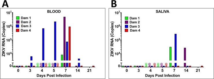
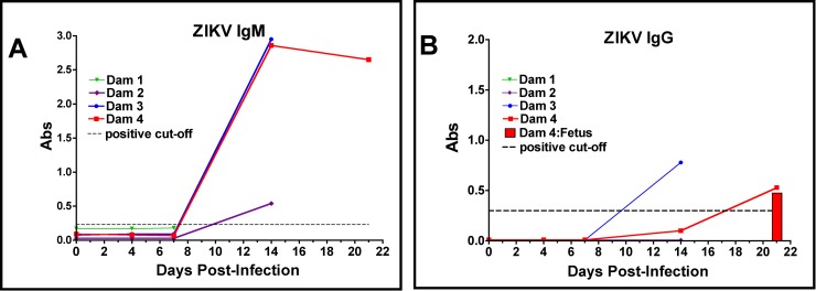
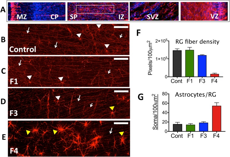
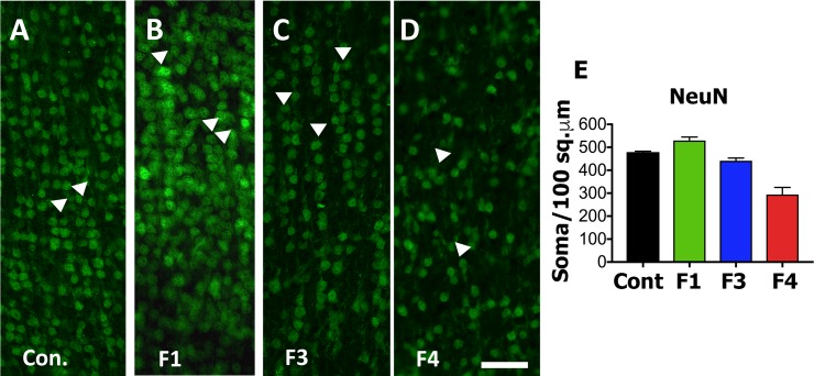
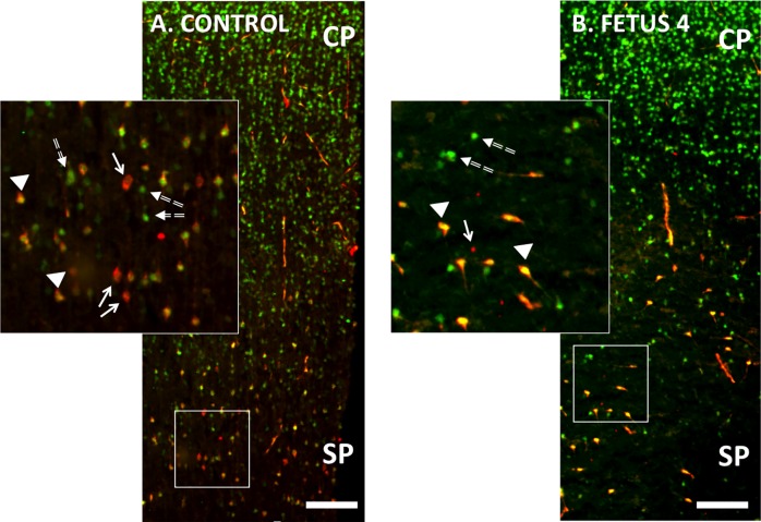
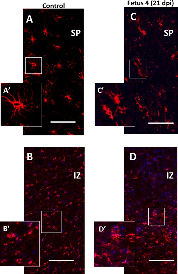
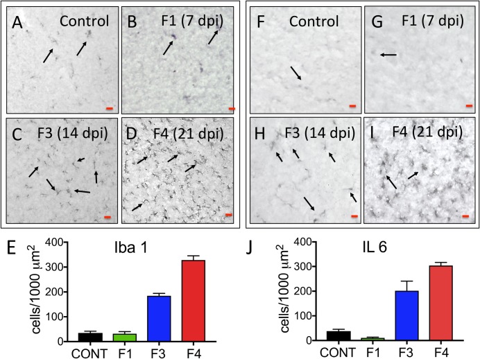
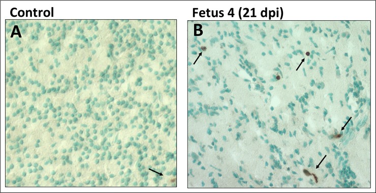
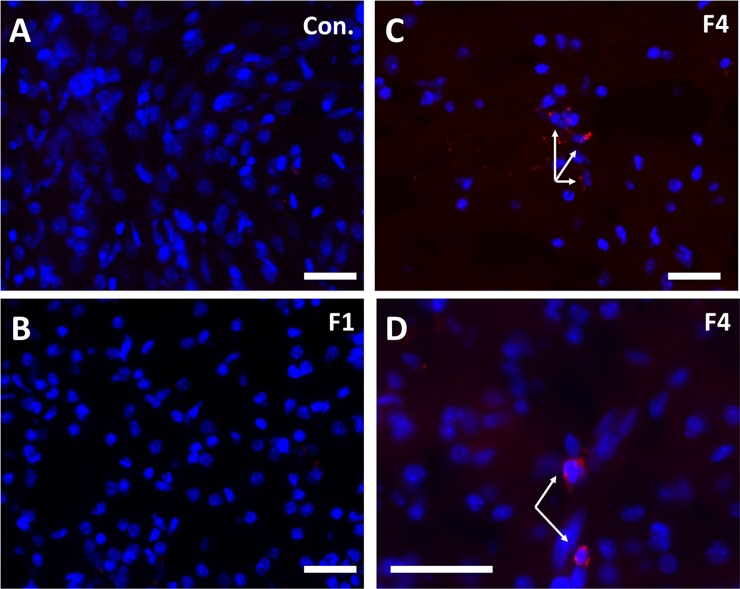
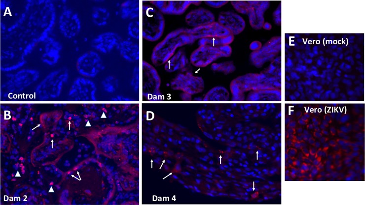
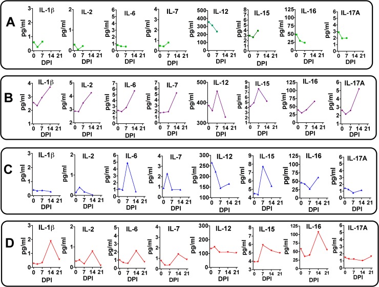
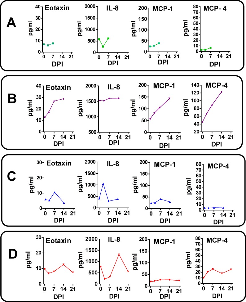
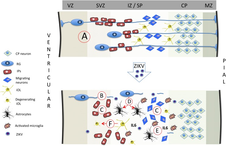
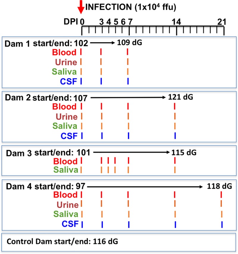
Zika virus (ZIKV) infection during pregnancy in humans is associated with an increased incidence of congenital anomalies including microcephaly as well as fetal death and miscarriage and collectively has been referred to as Congenital Zika Syndrome (CZS). Animal models for ZIKV infection in pregnancy have been developed including mice and non-human primates (NHPs). In macaques, fetal CZS outcomes from maternal ZIKV infection range from none to significant. In the present study we develop the olive baboon (Papio anubis), as a model for vertical transfer of ZIKV during pregnancy. Four mid-gestation, timed-pregnant baboons were inoculated with the French Polynesian ZIKV isolate (104 ffu). This study specifically focused on the acute phase of vertical transfer. Dams were terminated at 7 days post infection (dpi; n = 1), 14 dpi (n = 2) and 21 dpi (n = 1). All dams exhibited mild to moderate rash and conjunctivitis. Viremia peaked at 5-7 dpi with only one of three dams remaining mildly viremic at 14 dpi. An anti-ZIKV IgM response was observed by 14 dpi in all three dams studied to this stage, and two dams developed a neutralizing IgG response by either 14 dpi or 21 dpi, the latter included transfer of the IgG to the fetus (cord blood). A systemic inflammatory response (increased IL2, IL6, IL7, IL15, IL16) was observed in three of four dams. Vertical transfer of ZIKV to the placenta was observed in three pregnancies (n = 2 at 14 dpi and n = 1 at 21 dpi) and ZIKV was detected in fetal tissues in two pregnancies: one associated with fetal death at ~14 dpi, and the other in a viable fetus at 21 dpi. ZIKV RNA was detected in the fetal cerebral cortex and other tissues of both of these fetuses. In the fetus studied at 21 dpi with vertical transfer of virus to the CNS, the frontal cerebral cortex exhibited notable defects in radial glia, radial glial fibers, disorganized migration of immature neurons to the cortical layers, and signs of pathology in immature oligodendrocytes. In addition, indices of pronounced neuroinflammation were observed including astrogliosis, increased microglia and IL6 expression. Of interest, in one fetus examined at 14 dpi without detection of ZIKV RNA in brain and other fetal tissues, increased neuroinflammation (IL6 and microglia) was observed in the cortex. Although the placenta of the 14 dpi dam with fetal death showed considerable pathology, only minor pathology was noted in the other three placentas. ZIKV was detected immunohistochemically in two placentas (14 dpi) and one placenta at 21 dpi but not at 7 dpi. This is the first study to examine the early events of vertical transfer of ZIKV in a NHP infected at mid-gestation. The baboon thus represents an additional NHP as a model for ZIKV induced brain pathologies to contrast and compare to humans as well as other NHPs.


Conflict of interest statement

The authors have declared that no competing interests exist.

Figures

Fig 1
Fig 1
Viral loads (RNA) in whole blood (A) and saliva (B) from ZIKV-infected pregnant baboons. A one-step qRT-PCR was used to measure ZIKV RNA in whole blood (A) and saliva (B) from each animal at indicated days post-infection (copies per milliliter; ND: not determined).
Fig 2
Fig 2. Detection of anti-ZIKV antibody responses in pregnant baboon serum.
The presence of antibodies directed against ZIKV was determined by ELISA for IgM (A) or IgG (B). anti-ZIKV IgM were detected at 14 days post-infection in all three baboons sampled at this time point. Only two of the pregnant baboons had anti-ZIKV IgG; one at 14 days and the other by 21 days post-infection. The dam (2) with the weakest IgM response at 14 days did not exhibit an IgG response at this time point. The fetus of Dam 4 had anti-ZIKV IgG at levels similar to the mother indicating efficient FcγR transfer of IgG across the placenta.
Fig 3
Fig 3. Immunofluorescence (IF) for GFAP in the frontal cortex of fetal baboons from an uninfected control dam and from dams infected with ZIKV.
(A) Reconstruction of GFAP-IF in the frontal cortex of the fetal baboon (marginal zone (MZ), cortical plate (CP) subplate (SP), intermediate zone (IZ), subventricular zone (SVZ) and ventricular zone (VZ; (116 days gestation). B-E: GFAP-IF (RED) in the SP of the frontal cortex showing radial glial (RG) fibers (arrows) and occasional radial glia (white arrowheads) or astrocyte-appearing soma (yellow arrowheads) in control (B), and in three fetuses after maternal infection with ZIKV (C-E). Fetus 1 (C; 7 dpi; 109 days gestation) and Fetus 3 (D; 14 dpi; 115 days gestation) had an identical pattern of GFAP-IF RG fibers comparable to the control fetus. Neither Fetus 1 nor 3 had detectable ZIKV RNA in any fetal tissue including any of the cortical lobes. Fetus 4 (E; 21 dpi; 118 days gestation) that had detectable ZIKV RNA and protein in fetal frontal cortex had few continuous RG fibers (arrows). In the control fetus and Fetus 1, 2 and 3, occasional RG soma were noted (white arrowheads) with typically small soma and numerous radiating processes; In Fetus 3, an occasional larger soma consistent with astrocytes (yellow arrowheads) was noted. In Fetus 4, in addition to reduction in RG fibers, a notable dense astrocyte population was observed (yellow arrowheads). (F) Image analysis of astrocyte/radial glia soma (soma/100 μm2 of SP imaged per section, average ± SEM for three sections, 150 μm between sections) for each fetal frontal cortex. There was an ~3-fold increase in astrocytes/soma per unit area in the SP of Fetus 4 (F4) compared to the control fetus (Cont) or Fetus 1 (F1) or Fetus 3 (F3). (G) Image analysis of RG fiber density in the SP of each area imaged (GFAP+ fibers/100 μm2 of SP; average ± SEM for three sections per fetus) demonstrated that Fetus 4 (F4) had less than 10% of RG fibers remaining compared to the control fetus (Cont) or Fetus 1 or 3 (F1, F3). (bar = 25 μm).
Fig 4
Fig 4
NeuN immunofluorescence (Green) in the cortical plate of the frontal cortex of fetal baboons from an uninfected control dam (A), and from dams infected with ZIKV (B-D). Arrowheads indicate columns of NeuN labeled neurons in the CP of the control fetal cortex (A) and in the CP of Fetus 1 (B) and 3 (C) from ZIKV infected dams without viral transfer to the fetus. In contrast, Fetus 4 (D), which had detectable ZIKV RNA in the frontal cortex at 21 dpi had NeuN IF pattern that showed considerable disorganization consistent with the loss of radial glial fibers in the cortex of this fetus. Image analysis of NeuN+ neurons (E; NeuN+ cells/200 μm2 for three sections, mean ± SEM; 150 μm between sections) for each fetal frontal cortex (Control: Cont, Fetus 1: F1; Fetus 3: F3; Fetus 4: F4). There was a 2-fold decrease in NeuN+ IF neurons in the CP of the ZIKV infected cortex (Fetus 4) compared to the other fetal cortices. (bar = 25 μm).
Fig 5
Fig 5
Double immunofluorescence for NeuN (green; differentiating neurons) and Nestin (red; neuroprogenitors; NPCs) in the cortical plate (CP) and subplate (SP) of the developing frontal cortex of the control (A) and 21 day post ZIKV infection (B) fetal frontal cortex (Fetus 4). There were numerous differentiating neurons co-expressing NeuN and Nestin in the control cortex (arrows; yellow) as well as NPCs (Nestin: Red; arrowheads) and differentiated neurons (NeuN: Green; dashed arrows). In the ZIKV infected Fetus 4 (B), Nestin+ neurons (Green) appeared disorganized compared to the control cortex (which showed the characteristic pattern of columns of Nestin+ neurons migrating from the SP to the CP); only a few scattered remaining NPCs (NeuN: red) or differentiating neurons co-expressing NeuN and Nestin are observed in the ZIKV infected cortex. (bar = 100 μm).
Fig 6
Fig 6. Immunoflurorescence for O1 (immature oligodendrocytes; OL; Red) in the SP/IZ region of the developing fetal cortex.
In the control fetal frontal cortex SP (A: SP; B:IZ) there were numerous well developed immature oligodendrocytes (O1+; red) with extensive processes (see inset [A’]) while in the IZ region, the immature oligodendrocytes were smaller, had few processes and more abundant (B; B’: inset showing smaller, less differentiated oligodendrocytes). In the ZIKV infected Fetus 4 (C,D), the number of O1+ cells in the SP (C) and IZ (D) were similar but morphologically distinct with limited processes in the SP and in both SP and IZ, the immature oligodendrocytes had the appearance of degeneration or arrested development. (bar = 50 μm).
Fig 7
Fig 7
Immunohistochemistry for neuroinflammatory markers (microglia: Iba1; A-D; Interleukin 6 [IL-6]; F-I) in the frontal cortex of control and fetuses from dams infected with ZIKV. In both control (A,F) and Fetus 1 (7 dpi; B,G), occasional Iba1 reactive microglia and IL-6+ cells were observed (arrows). In the 14 dpi Fetus 3 (C,H) an increase in the number of Iba1+ microglia and IL-6 immunostaining cells were observed throughout the IZ through the CP (E), despite the fetal cortex being negative for ZIKV RNA. The 21 dpi Fetus 4 (D,I) that was ZIKV RNA+, exhibited the greatest Iba1+ microglia and IL-6 immunostaining (J). (For E, J: IF+ cells/200 μm2; mean ± SEM for three sections per fetus; 150 μm between sections).
Fig 8
Fig 8
TUNEL staining for apoptosis in the frontal cortex of the control (A) and 21 day post-infection Fetus 4 (B). The cortex of the control fetus had very few apoptotic cells compared to the ZIKV infected fetus (brown cells; arrows). While there were dispersed TUNEL stained cells in the infected fetus, the density was not consistent with widespread apoptosis in the cortex of the infected fetus at 21 days post-infection.
Fig 9
Fig 9
Pan flavivirus immunofluorescence (IF; Red: flavivirus; Blue: DAPI) staining in subplate/intermediate zone in the control fetus (A), 7 day post-ZIKV infection Fetus 1 (B) with no detection of virus in fetal tissues (B) and Fetus 4 at 21 days post-ZIKV infection that was ZIKV RNA+ in the frontal cortex (C,D). There was no IF for ZIKV in frontal cortex in either control (A) or 7 dpi fetus (B). Scattered ZIKV+ IF (Red) was detected in the SP/IZ of fetus 4 that was primarily perinuclear in localization (C 10x, D, 20x objective magnification; bar = 25μm).
Fig 10
Fig 10
Pan flavivirus immunofluorescence (IF; Red: flavivirus; Blue: DAPI) staining in placenta from control (A), and three placenta from ZIKV infected dams that ZIKV RNA+ in placenta (B, Dam 3, 14 dpi; C, Dam 3, 14 dpi; D, Dam 4, 21 dpi). The most intense IF was observed in the placenta of Dam 2 (B) in which there was fetal death. ZIKV IF was observed in syncytiotrophoblast (arrows) in Dam 2 and 3. Arrowheads in B indicate IF in the villous cores which could be either stromal or endothelial cells. A less intense IF pattern was observed in Dam 4 (D) in the overlying syncytiotrophoblast which is covering the underlying cytotrophoblasts, despite widespread vertical transfer of ZIKV to the fetus.
Fig 11
Fig 11. Maternal plasma cytokine concentrations in response to ZIKV infection in timed pregnant baboons.
Only cytokines that were detectable and exhibited changes in concentration in response to ZIKV infection are shown. In Dam 1 (A) studied for 7 days post-infection, no chemokines increased post-infection. For Dam 2 (B), increases in IL-β, IL-2, IL-7, IL-12, IL-15, IL-16 and IL-17A were observed with peak levels either at day 7 or 14 post-infection. For Dam 3 (C) acute (day 7) increases were observed for IL-6, IL-7 and IL-15 (with a small increase at day 7 for IL-2). For Dam 4 (D) increases in IL-1β, IL-2, IL-6 IL-7, IL-15 and IL-16 were observed with peak levels either at day 14 (exception IL-15 at day 14), with resolution by study termination at day 21 for this dam.
Fig 12
Fig 12. Maternal plasma chemokine concentrations in response to ZIKV infection in timed pregnant baboons.
Only chemokines that were detectable and exhibited changes in concentration in response to ZIKV infection are shown. In Dam 1 (A) studied for 7 days post-infection, no chemokines increased post-ZIKV infection. For Dam 2 (B), increases in Eotaxin, MCP-1 and MCP-4 were observed peaking at day 14 post-infection. For Dam 3 (C) a transient increase in Eotaxin and IL-8 was observed. For Dam 4 (D), a similar transient increase in Eotaxin, IL-8 and MCP-4 was noted.
Fig 13
Fig 13. Summary diagram depicting potential mechanisms via which ZIKV may lead to fetal brain pathology in primates.
(A) In the normally developing primate cortex, radial glial (RG) fibers serve as scaffolding for migrating neurons and intermediate precursors (IP) to the CP [55]. During the second half of gestation, RG end-feet detach from the ventricular and pial surfaces removing the RG fiber scaffolding after the cortex has formed its layers [–59]. During the second half of gestation, RG also gradually differentiate to astrocytes and pre-oligodendrocytes [–61] with normal transformation to astrocytes in the fetal frontal cortex. Based on the present study and studies in macaques (33,34,37,38), ZIKV infection of the fetal cortex results in a loss in RG fibers (B) resulting in disorganized neuroprogenitor cells (C; intermediate precursors (IP) and migrating neurons). (D) The widely reported astrogliosis may be the result of premature differentiation of RG to astrocytes, and/or as part of the noted neuroinflammatory response to ZIKV that includes increased microglia and IL-6 (E). (F) ZIKV infection impacts developing oligodendrocytes leading to reduced myelination as observed in the pigtail macaque and as reported in human fetuses [37, 38]. (Abbreviations: VZ: ventricular zone; SVZ: subventricular zone; IZ: intermediate zone; SP: subplate; CP: cortical plate; MZ: marginal zone).
Fig 14
Fig 14. Schematic representation of the experimental design.
Multiparous timed pregnant olive baboons (n = 4) were infected subcutaneously (1x10⁴ ffu, 1 ml volume, strain H/FP/2103) on Day 0. Maternal blood, urine, CSF and saliva were obtained on the indicated days for each animal. The gestational age at the time of infection ranged from 97–107 days gestation (dG). One dam was euthanized at day 7 post infection (Dam 1), two dams on day 14 post infection (Dams 2,3) and on at 21 days post infection for collection of maternal and fetal tissues. The gestational age range at necropsy was 109–121 dG; a timed pregnant control dam was euthanized at 116 dG.
Fig 15
Fig 15. 120 day gestation fetal baboon brain depicting the lobes and location of the sections used for immunohistochemical analysis (white line in frontal cortex).

References

    1. Dick GW. Zika virus. II. Pathogenicity and physical properties. Transactions of the Royal Society of Tropical Medicine and Hygiene. 1952;46(5):521–34. Epub 1952/09/01. . - PubMed
    1. Dick GW, Kitchen SF, Haddow AJ. Zika virus. I. Isolations and serological specificity. Transactions of the Royal Society of Tropical Medicine and Hygiene. 1952;46(5):509–20. Epub 1952/09/01. . - PubMed
    1. Brasil P, Pereira JP Jr., Moreira ME, Ribeiro Nogueira RM, Damasceno L, Wakimoto M, et al. Zika Virus Infection in Pregnant Women in Rio de Janeiro. N Engl J Med. 2016;375(24):2321–34. 10.1056/NEJMoa1602412 - DOI - PMC - PubMed
    1. Chan JF, Choi GK, Yip CC, Cheng VC, Yuen KY. Zika fever and congenital Zika syndrome: An unexpected emerging arboviral disease. J Infect. 2016;72(5):507–24. Epub 2016/03/05. 10.1016/j.jinf.2016.02.011 . - DOI - PMC - PubMed
    1. Alvarado MG, Schwartz DA. Zika Virus Infection in Pregnancy, Microcephaly, and Maternal and Fetal Health: What We Think, What We Know, and What We Think We Know. Arch Pathol Lab Med. 2017;141(1):26–32. Epub 2016/09/17. 10.5858/arpa.2016-0382-RA . - DOI - PubMed

Publication types

MeSH terms