Review
. 2019 Mar 1;43(2):145-161.
doi: 10.1093/femsre/fuy045.

Microbial uptake by the respiratory epithelium: outcomes for host and pathogen

Affiliations
Review

Microbial uptake by the respiratory epithelium: outcomes for host and pathogen

Margherita Bertuzzi et al. FEMS Microbiol Rev. .

Abstract

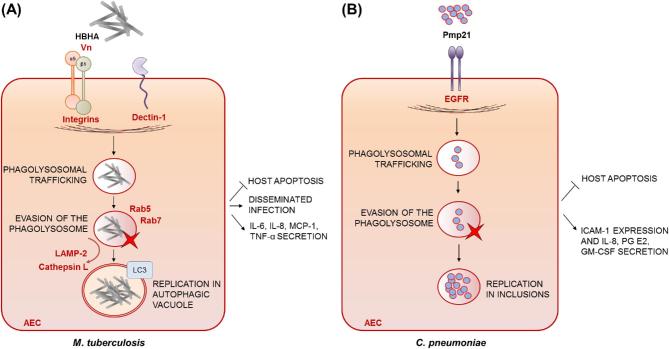
Intracellular occupancy of the respiratory epithelium is a useful pathogenic strategy facilitating microbial replication and evasion of professional phagocytes or circulating antimicrobial drugs. A less appreciated but growing body of evidence indicates that the airway epithelium also plays a crucial role in host defence against inhaled pathogens, by promoting ingestion and quelling of microorganisms, processes that become subverted to favour pathogen activities and promote respiratory disease. To achieve a deeper understanding of beneficial and deleterious activities of respiratory epithelia during antimicrobial defence, we have comprehensively surveyed all current knowledge on airway epithelial uptake of bacterial and fungal pathogens. We find that microbial uptake by airway epithelial cells (AECs) is a common feature of respiratory host-microbe interactions whose stepwise execution, and impacts upon the host, vary by pathogen. Amidst the diversity of underlying mechanisms and disease outcomes, we identify four key infection scenarios and use best-characterised host-pathogen interactions as prototypical examples of each. The emergent view is one in which effi-ciency of AEC-mediated pathogen clearance correlates directly with severity of disease outcome, therefore highlighting an important unmet need to broaden our understanding of the antimicrobial properties of respiratory epithelia and associated drivers of pathogen entry and intracellular fate.

Keywords: airway epithelial cells (AECs); epithelial responses; microbial uptake; microbicidal activities; pathogenesis; respiratory epithelium.


Figures

Figure 1.
Figure 1.
Microbial uptake by AECs occurs in a step wise manner and leads to species-specific outcomes for both host and pathogen. Receptor- or effector-mediated uptake of microbes by AECs leads to the activation of host cellular signalling pathways and remodelling of the host cytoskeleton at the site of pathogen entry. A successful antimicrobial defence results from trafficking of the internalised pathogen, through a series of intracellular compartments culminating in phagolysosome-mediated killing. In some cases however, intracellular pathogens evade intracellular killing either by thriving in the extremely hostile environment of the AEC phagolysosome or by preventing the fusion of the microbe-containing vacuole with lysosomes. By residing latently in the intracellular niche or replicating, such pathogens successfully exploit internalisation by AECs to cause host damage and trigger inflammatory responses, ultimately leading to disseminated infection.
Figure 2.
Figure 2.
Microbial uptake leading to direct neutralisation of pathogen. (A) Burkholderia cepacia complex: once internalised by wild-type AECs, species of the B. cepacia complex (Bcc) are trafficked to cathepsin D-positive endocytic vesicles and killed. Uptake by AECs occurs in a CFTR-dependent manner and via an uncharacterised glycolipid receptor and requires Bcc lipases, the flagellum, cable pilin and the 22-kDa adhesin protein, which binds to the host surface protein cytokeratin 13 (CK13). Exogenous addition of IL-8 enhances intracellular bacterial growth. (B) Aspergillus fumigatus spores: following uptake by AECs, the majority of internalised A. fumigatus spores are killed. Aspergillus fumigatus uptake is mediated by E-cadherin and CFTR, by Dectin-1 via binding of fungal β-glucan and α5β1 integrin via binding of A. fumigatus CalA. The gliotoxin immunotoxin also facilitates spore internalisation by AECs. (C) Capsule-deficient K. pneumoniae variants: upon uptake by AECs, capsule-deficient K. pneumoniae are efficiently killed. Klebsiella pneumoniae is internalised by AECs in a process that involves a GlcNAc-binding surface component and an N-glycosylated receptor on the host cell surface. Also, AEC-mediated C3 opsonisation enhances dramatically CD46-mediated microbial uptake. Klebsiella pneumoniae uptake increases surface expression of ICAM-1 and secretion of IL-8 by AECs in an NF-kB-dependent manner. Pathogen-derived effectors of uptake are indicated in bold black font, putative or proven host receptors, opsonins or bridging factors driving uptake are indicated in bold red font.
Figure 3.
Figure 3.
Microbial clearance facilitated by cellular desquamation and apoptosis of infected AECs: P. aeruginosa. (A) In healthy AECs, internalisation of P. aeruginosa leads to initiation of NF-κB nuclear translocation, cellular desquamation and eventual apoptosis and shedding of the infected cells. Intracellular P. aeruginosa viability is not reduced within the host cell and P. aeruginosa replicates in plasma membrane blebs (PMBs) via products secreted by a bacterial type III secretion system. Pseudomonas aeruginosa uptake is dependent on the bacterial lipopolysaccharide (LPS)–core oligosaccharide and CFTR and, in a strain-dependent manner, on αvβ5 and α5β1integrins via vitronectin (Vn) and fibronectin (Fn) bridging, respectively. The interaction of the two major bacterial adhesion factors, namely type IV (Tfp) and flagella, with the N-glycoproteins and heparate sulfate proteoglycans (HSPG), respectively, is also required for microbial uptake, as well as the effector proteins of the secretion systems H2-T6SS and H3-T6SS. Internalisation-mediated apoptosis limits the release of cytokines, such as IL-1β. (B) Following uptake of P. aeruginosa, CF epithelial cells undergo significantly delayed apoptosis compared with wild-type AECs allowing higher rates of intracellular replication and sustained host damage. Pathogen-derived effectors of uptake are indicated in bold black font, putative or proven host receptors, opsonins or bridging factors driving uptake are indicated in bold red font.
Figure 4.
Figure 4.
Microbial uptake leading to incomplete killing, host damage and disseminated infection. (A) Staphylococcus aureus: upon uptake by AECs, S. aureus is able to undergo intracellular replication within, and cause apoptosis of, cultured AECs to further disseminate and cause disease. EsxA and EsxB, members of the ESAT-6-like secretion system, contribute to bacterial release from host cells. Staphylococcus aureus uptake by AECs occurs via the interaction of FnBPs, FN-binding proteins, and the host integrin α5β1. The extracellular adherence protein Eap also binds to Fn, while FnBPs bind the heat shock protein 60 (Hsp60). (B) Streptococcus pneumoniae: upon uptake by AECs, internalised S. pneumoniae co-localises with several endosomal markers such as LAMP-1, Rab5, Rab4 and Rab7, indicating bacterial intracellular trafficking to recycling endosomes for transcytosis and/or killing in phagolysosomes. However, complete killing is not achieved and remaining pneumococci may persist, without multiplying, leading to transcytosis. Streptococcus pneumoniae αvβ3 integrin internalisation by AECs is mediated by Vn, the plasminogen and Fn-binding protein PepO and the choline-binding protein CbpA/PspC, which binds to the complement regulator factor H and the human polymeric immunoglobulin receptor (pIgR). AECs infected with S. pneumoniae release IL-8 in an internalisation-dependent manner. (C) Haemophilus influenzae: NTHI variants can become internalised by AECs to subsequently reside intracellularly and trigger apoptosis of the infected cells. Integrins, Dectin-1, CEACAMs, PAFr and Vn are essential for H. influenzae uptake by AECs. Internalisation via PAFr occurs via the interaction with ChoP of the lipooligosaccharide (LOS), while surface fibrils on NTHIs allow the interaction with Vn. Pathogen-derived effectors of uptake are indicated in bold black font, putative or proven host receptors, opsonins or bridging factors driving uptake are indicated in bold red font.
Figure 5.
Figure 5.
Microbial uptake leading to intracellular survival of pathogen and AEC apoptosis. (A) Mycobacterium tuberculosis: once internalised by AECs, M. tuberculosis is trafficked to late endosomes (Rab5 and Rab7 positive) but no co-localisation with the lysosomal markers LAMP-2 and cathepsin L is observed. Instead, M. tuberculosis is diverted to the autophagy pathway, providing the means to evade intracellular killing. Mycobacterium tuberculosis is able to proliferate intracellularly within AECs and safeguard its replicative niche by suppressing epithelial apoptosis. β1 and αν integrin act synergistically in mediating uptake by AECs as do an unidentified Vn receptor and Dectin-1. The heparin-binding haemagglutinin (HBHA) is the main M. tuberculosis surface component interacting with AECs, but its cognate receptor remains unidentified. Mycobacterium tuberculosis uptake stimulates the release of IL-8, IL-6, monocyte chemotactic protein-1 (MCP-1) and tumour necrosis factor α (TNF-α) in a process dependent on intracellular bacterial growth and Dectin-1. (B) Chlamydia pneumoniae: following uptake by AECs, C. pneumoniae occupies membrane-bound vacuoles called inclusions, and inhibits apoptosis of infected cells. The bacterial invasin Pmp21 drives internalisation into AECs via binding to the human epithermal growth factor receptor (EGFR). Chlamydia pneumonia uptake by AECs induces the release of IL-8, prostaglandin E2 and granulocyte macrophage colony-stimulating factor (GM-CSF), as well as an increase in the expression of ICAM-1. Pathogen-derived effectors of uptake are indicated in bold black font, putative or proven host receptors, opsonins or bridging factors driving uptake are indicated in bold red font.

Similar articles

Cited by

References

    1. Aderem A, Underhill DM. Mechanisms of phagocytosis in macrophages. Annu Rev Immunol 1999;17:593–623. - PubMed
    1. Agarwal V, Ahl J, Riesbeck K et al. .. An alternative role of C1q in bacterial infections: facilitating Streptococcus pneumoniae adherence and invasion of host cells. J Immunol 2013a;191:4235–45. - PubMed
    1. Agarwal V, Asmat TM, Luo S et al. .. Complement regulator Factor H mediates a two-step uptake of Streptococcus pneumoniae by human cells. J Biol Chem 2010;285:23486–95. - PMC - PubMed
    1. Agarwal V, Hammerschmidt S. Cdc42 and the phosphatidylinositol 3-kinase-Akt pathway are essential for PspC-mediated internalization of pneumococci by respiratory epithelial cells. J Biol Chem 2009;284:19427–36. - PMC - PubMed
    1. Agarwal V, Kuchipudi A, Fulde M et al. .. Streptococcus pneumoniae endopeptidase O (PepO) is a multifunctional plasminogen- and fibronectin-binding protein, facilitating evasion of innate immunity and invasion of host cells. J Biol Chem 2013b;288:6849–63. - PMC - PubMed

Publication types