Review
. 2019 Mar 1;160(3):626-638.
doi: 10.1210/en.2018-00794.

Epithelial Mutations in Endometriosis: Link to Ovarian Cancer

Affiliations
Review

Epithelial Mutations in Endometriosis: Link to Ovarian Cancer

Serdar E Bulun et al. Endocrinology. .

Abstract

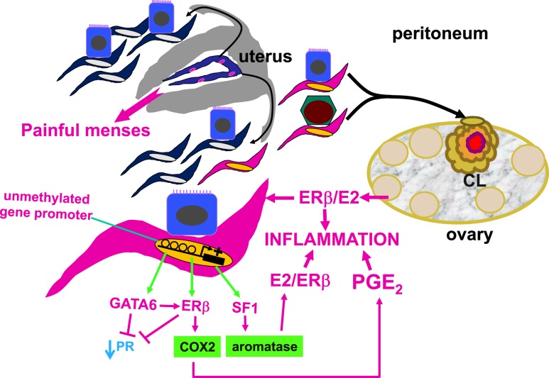
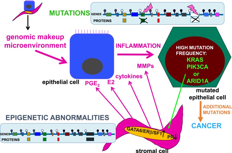
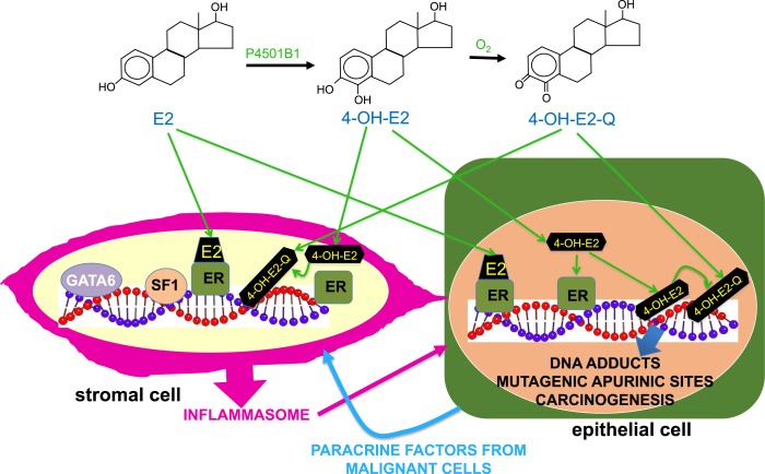
Epidemiologic and histopathologic associations between endometriosis and epithelial ovarian cancer have been reported; however, the underlying molecular and cellular mechanisms are not well understood. A possible genetic link has been suggested in recent publications. Driver mutations in PIK3CA, KRAS, ARID1A, and other genes have been found in the epithelium of intrauterine endometrial tissue, ovarian and extraovarian pelvic endometriosis tissue, ovarian cancers associated with endometriosis (i.e., clear cell and endometrioid type), and other epithelial ovarian cancers. This makes sense because pelvic endometriosis occurs primarily as a result of retrograde menstruation and implantation of endometrial tissue fragments in ovarian inclusion cysts or extraovarian peritoneal or subperitoneal sites. Unlike epithelial cells, endometriotic stromal cells are mutation free but contain widespread epigenetic defects that alter gene expression and induce a progesterone-resistant and intensely inflammatory environment, driven by estrogen via estrogen receptor-β. The resulting increased estrogenic action in the stroma drives inflammation and sends paracrine signals to neighboring epithelial cells to enhance proliferation. In addition, massively high concentrations of estrogen in the ovary may exert an additional and direct genotoxic effect on DNA and cause accumulation of additional mutations and malignant transformation in initially mutated endometriotic epithelial cells in an ovarian endometrioma, which may initiate epithelial ovarian cancer. The same epithelial mutations and inflammatory processes in stroma are seen in extraovarian deep-infiltrating endometriosis, but carcinogenesis does not occur. We provide a focused review of the literature and discuss the implications of recent genetic breakthroughs linking endometriosis and ovarian cancer.


Figures

Figure 1.
Figure 1.
Central roles of the endometrial stromal cell and epigenetic regulation in endometriosis. In a nonpregnant ovulatory woman, endometrium is shed and renewed every month. Although most of the menstrual material composed of endometrial tissue fragments is expelled through the cervix into the vagina, a portion of it travels retrograde into the lower abdominal cavity and spills on pelvic tissues, including the ovaries. Although retrograde menstruation is observed in most women, only ∼10% of premenopausal women develop the histologic evidence or symptoms of endometriosis. Histologically, the majority of pelvic endometriotic lesions contain endometrial stromal cells, whereas fewer lesions harbor epithelial cells that are relatively scanty. If these endometrial tissue fragments implant on the peritoneum that covers the pelvic portions of the rectovaginal pouch, bowel, uterus, or pelvic side walls, these lesions are usually referred to as peritoneal endometriosis. If they line up along an ovarian inclusion cyst wall, this arrangement eventually evolves into an ovarian endometrioma, which, over time, accumulates remarkable amounts of blood and inflammatory products. Results of molecular and cellular studies suggest the eutopic endometrium of women with endometriosis contains stem-like stromal cell types that travel to the pelvis via retrograde menstruation. These stromal cells exhibit widespread epigenetic defects, such as inappropriately unmethylated DNA at gene promoters. This leads to inappropriate expression of the transcription factors, GATA6, ERβ, and SF1. These critical factors initiate a vicious cascade that activates the expression of cyclooxygenase-2 and aromatase, which leads to the production of large quantities of E2 and PGE2 fueling inflammation. GATA6 and ERβ suppress PR expression, causing progesterone resistance. If the endometrial tissue fragments become trapped in a CL cyst that has recently ruptured, they may survive and give rise to an ovarian endometrioma. CL, corpus luteum; E2, estradiol; ERβ, estrogen receptor-β; GATA6, GATA-binding factor-6; PGE2, prostaglandin E2; PR, progesterone receptor; SF1, steroidogenic factor-1.
Figure 2.
Figure 2.
Interactions between the genomic processes in endometriotic stromal and epithelial cells. The genomic composition and microenvironment (intraovarian vs extraovarian) determine the malignant transformation of endometriotic lesions. The stromal cells do not contain mutations but do show abnormal epigenetic marks that modulate gene expression. For example, DNA methylation (closed circles) or unmethylation (open circles) silences or permits transcription of specific genes. In endometriotic stromal cells, progesterone resistance is due to suppression of PR and overexpression of GATA6, ERβ, and SF1 proteins. Inflammatory factors and proteins that remodel endometrial tissues, such as PGE2, E2, cytokines, and MMPs, accumulate in the stroma. In endometriotic epithelial cells, tumor driver mutations disrupt critical protein function, including PIK3CA, KRAS, ARID1A, and many others. The highly inflammatory and estrogenic, as well as progesterone-resistant, microenvironment in ovarian endometriomas may enhance the accumulation of additional mutations and proliferation of epithelial cells, which eventually become malignant and invasive. The specific effects of these epithelial mutations of stromal cell function in endometrial or endometriotic tissue are currently unknown. MMP, matrix metalloproteinase; PGE2, prostaglandin E2.
Figure 3.
Figure 3.
Ovarian microenvironment, mutagenesis, and carcinogenesis. Endometriotic epithelial cells with driver mutations in extraovarian sites do not seem to become malignant, whereas identical driver mutations are found in endometriosis-associated ovarian cancer, suggesting that the ovarian microenvironment is unique for enabling initially mutated endometriotic epithelial cells to acquire additional mutations and become malignant. In ovarian tissue, E2 and other estrogens are produced at levels that are >10,000 times those present in peripheral blood. We hypothesize that the genotoxic metabolites of estrogens cause DNA adducts resulting in mutagenic apurinic sites and accumulation of additional mutations during cell division. In fact, the levels of the CYP1B1 enzyme in endometriotic stromal cells are ∼100 times those found in normal endometrial stromal cells. This enzyme catalyzes the conversion of E2 to its 4-hydroxy-catechol metabolite, 4-OH-E2, which is further converted to redox-active quinones (4-OH-E2-Q). 4-OH-E2-Q may cause DNA damage by alkylation or oxidation leading to mutagenesis. Accumulation of more mutations in addition to an original driver mutation (see Fig. 2) likely facilitates the malignant transformation of an endometriotic epithelial cell located in an ovarian inclusion cyst. These epithelial cells lie adjacent to mutation-free but epigenetically defective stromal cells that produce inflammatory substances under the control of several transcription factors, including GATA6, SF1, and ERβ. In addition to regulating gene transcription, both ERα and ERβ in epithelial cells may concentrate toxic estrogen metabolites at DNA sites to intensify DNA damage and mutagenesis.

References

    1. Bulun SE. Endometriosis In: Strauss J, Narnieri R, eds. Yen & Jaffe’s Reproductive Endocrinology. Philadelphia, PA: Elsevier; 2018:609–642.
    1. Anglesio MS, Papadopoulos N, Ayhan A, Nazeran TM, Noë M, Horlings HM, Lum A, Jones S, Senz J, Seckin T, Ho J, Wu RC, Lac V, Ogawa H, Tessier-Cloutier B, Alhassan R, Wang A, Wang Y, Cohen JD, Wong F, Hasanovic A, Orr N, Zhang M, Popoli M, McMahon W, Wood LD, Mattox A, Allaire C, Segars J, Williams C, Tomasetti C, Boyd N, Kinzler KW, Gilks CB, Diaz L, Wang TL, Vogelstein B, Yong PJ, Huntsman DG, Shih IM. Cancer-associated mutations in endometriosis without cancer. N Engl J Med. 2017;376(19):1835–1848. - PMC - PubMed
    1. Li X, Zhang Y, Zhao L, Wang L, Wu Z, Mei Q, Nie J, Li X, Li Y, Fu X, Wang X, Meng Y, Han W. Whole-exome sequencing of endometriosis identifies frequent alterations in genes involved in cell adhesion and chromatin-remodeling complexes. Hum Mol Genet. 2014;23(22):6008–6021. - PubMed
    1. Suda K, Nakaoka H, Yoshihara K, Ishiguro T, Tamura R, Mori Y, Yamawaki K, Adachi S, Takahashi T, Kase H, Tanaka K, Yamamoto T, Motoyama T, Inoue I, Enomoto T. Clonal expansion and diversification of cancer-associated mutations in endometriosis and normal endometrium. Cell Reports. 2018;24(7):1777–1789. - PubMed
    1. Sampson JA. Peritoneal endometriosis due to the menstrual dissemination of endometrial tissue into the peritoneal cavity. Am J Obstet Gynecol. 1927;14(4):422–469.

Publication types