. 2019 Oct 1;111(10):1068-1077.
doi: 10.1093/jnci/djz005.

Targeted Genomic Profiling of Acral Melanoma

Targeted Genomic Profiling of Acral Melanoma

Iwei Yeh et al. J Natl Cancer Inst. .

Abstract

Background: Acral melanoma is a rare type of melanoma that affects world populations irrespective of skin color and has worse survival than other cutaneous melanomas. It has relatively few single nucleotide mutations without the UV signature of cutaneous melanomas, but instead has a genetic landscape characterized by structural rearrangements and amplifications. BRAF mutations are less common than in other cutaneous melanomas, and knowledge about alternative therapeutic targets is incomplete.

Methods: To identify alternative therapeutic targets, we performed targeted deep-sequencing on 122 acral melanomas. We confirmed the loss of the tumor suppressors p16 and NF1 by immunohistochemistry in select cases.

Results: In addition to BRAF (21.3%), NRAS (27.9%), and KIT (11.5%) mutations, we identified a broad array of MAPK pathway activating alterations, including fusions of BRAF (2.5%), NTRK3 (2.5%), ALK (0.8%), and PRKCA (0.8%), which can be targeted by available inhibitors. Inactivation of NF1 occurred in 18 cases (14.8%). Inactivation of the NF1 cooperating factor SPRED1 occurred in eight cases (6.6%) as an alternative mechanism of disrupting the negative regulation of RAS. Amplifications recurrently affected narrow loci containing PAK1 and GAB2 (n = 27, 22.1%), CDK4 (n = 27, 22.1%), CCND1 (n = 24, 19.7%), EP300 (n = 20, 16.4%), YAP1 (n = 15, 12.3%), MDM2 (n = 13, 10.7%), and TERT (n = 13, 10.7%) providing additional and possibly complementary therapeutic targets. Acral melanomas with BRAFV600E mutations harbored fewer genomic amplifications and were more common in patients with European ancestry.

Conclusion: Our findings support a new, molecularly based subclassification of acral melanoma with potential therapeutic implications: BRAFV600E mutant acral melanomas with characteristics similar to nonacral melanomas that could benefit from BRAF inhibitor therapy, and non-BRAFV600E mutant acral melanomas. Acral melanomas without BRAFV600E mutations harbor a broad array of therapeutically relevant alterations. Expanded molecular profiling would increase the detection of potentially targetable alterations for this subtype of acral melanoma.


Figures

Figure 1.
Figure 1.
Spectrum of MAPK activating genetic alterations in acral melanoma. Each column represents a single sample (n = 122). Each row indicates reportable findings for each gene(s) as designated by the legend. Many samples have multiple reportable findings.
Figure 2.
Figure 2.
KIT mutations in acral melanoma. A) KIT mutations are distributed between the juxtamembranous (JMD) and kinase domains. B) Activating mutations occur in various exons of KIT and in some cases, the mutant KIT allele is amplified. C) KIT amplification often affects flanking genes PDGFRA and KDR. Details of the chromosome 4q12 amplicon showing log2 scaled copy number ratios in KIT amplified cases.
Figure 3.
Figure 3.
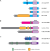
Fusion kinases in acral melanoma. A) The BRAF fusion junctions reside downstream of the autoinhibitory RAS binding domain (RBD) and upstream of the kinase domain of BRAF. ERC1-BRAF contains a coiled coil domain that promotes dimerization is contributed by ERC1. B) The predicted NTRK3 fusion proteins are missing most of the extracellular domain of NTRK3 and may contain the transmembrane domain in addition to the kinase domain. The MYO5A-NTRK3 fusion contains coiled-coil domains contributed by MYO5A. C) The predicted ATP2B4-PRKCA fusion protein lacks the regulatory calcium binding domains (C1, C2, C3).
Figure 4.
Figure 4.
NF1 and SPRED1 loss in acral melanoma. A) NF1 immunohistochemistry in an acral melanoma without NF1 mutation (top) shows robust expression of NF1 within neoplastic melanocytes, whereas as expression is lost in a melanoma with a truncating mutation in NF1 and loss of the wild-type allele (bottom, residual staining is in endothelial cells). B) NF1 and SPRED1 loss are mutually exclusive. All cases with bi-allelic loss of NF1 or SPRED1 (deep deletion or loss of function mutation with loss of the wild-type allele) and/or absence of NF1 protein expression by immunohistochemistry are shown, one sample per column. Additional MAPK pathway activating alterations are present in a majority of cases.
Figure 5.
Figure 5.
Alterations affecting the CDK4/6 pathway in acral melanoma. A) In this tiling plot, each column represents a single sample. Each row indicates reportable findings for each gene(s) as designated by the legend. Many samples have multiple reportable findings. B) A pathway diagram shows the interactions of the components of the CDK4/6 pathway. C) p16 immunohistochemistry demonstrates loss of expression in cases without bi-allelic inactivation. Acral melanomas with shallow deletion of CDKN2A (corresponding to heterozygous loss) demonstrate variable p16 levels by immunohistochemistry. In a case (KAM130) with shallow deletion of CDKN2A, strong cytoplasmic and nuclear p16 is present in neoplastic melanocytes (top panel). In another case (KAM34) with shallow deletion of CDKN2A, neoplastic melanocytes are negative for p16 by immunohistochemistry (bottom panel). Nuclear p16 positivity is present within endothelial cells.
Figure 6.
Figure 6.
Alterations affecting the p53 pathway in acral melanoma. A) In this tiling plot, each column represents a single sample. Each row indicates reportable findings for each gene(s) as designated by the legend. Many samples have multiple reportable findings. B) A pathway diagram shows the interactions of the components of the p53 pathway. C) Copy number profile of chromosome 12 shows high level amplification of MDM2 at a higher level than CDK4 (case MB_1424). D) Left: Whole genome copy number profile shows multiple copy number gains and losses and high level amplification of EP300 (KAM5). Right: Higher resolution view of EP300 amplification on chromosome 22.

References

    1. Desai A, Ugorji R, Khachemoune A.. Acral melanoma foot lesions. Part 1: epidemiology, aetiology, and molecular pathology. Clin Exp Dermatol. 2017;428:845–848. - PubMed
    1. Durbec F, Martin L, Derancourt C, Grange F.. Melanoma of the hand and foot: epidemiological, prognostic and genetic features. A systematic review. Br J Dermatol. 2012;1664:727–739. - PubMed
    1. Bello DM, Chou JF, Panageas KS.. Prognosis of acral melanoma: a series of 281 patients. Ann Surg Oncol. 2013;2011:3618–3625. - PubMed
    1. Turajlic S, Furney SJ, Lambros MB, et al. Whole genome sequencing of matched primary and metastatic acral melanomas. Genome Res. 2012;222:196–207. - PMC - PubMed
    1. Curtin JA, Fridlyand J, Kageshita T, et al. Distinct sets of genetic alterations in melanoma. N Engl J Med. 2005;35320:2135–2147. - PubMed

Publication types

MeSH terms

Substances