. 2019 May:125:146-153.
doi: 10.1016/j.nbd.2019.01.004. Epub 2019 Jan 15.

Long-term RNAi knockdown of α-synuclein in the adult rat substantia nigra without neurodegeneration

Affiliations

Long-term RNAi knockdown of α-synuclein in the adult rat substantia nigra without neurodegeneration

Alevtina Zharikov et al. Neurobiol Dis. 2019 May.

Abstract

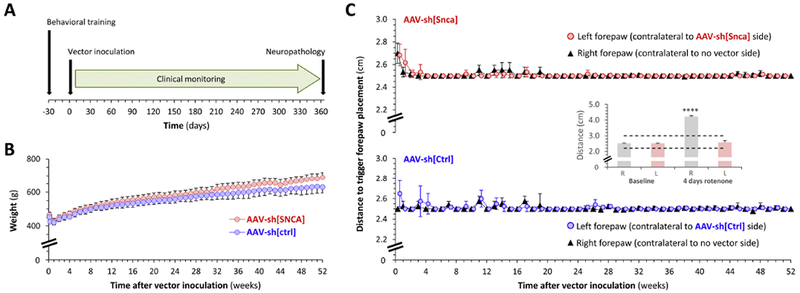
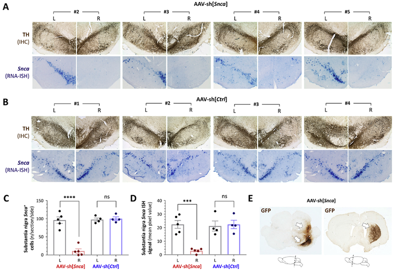
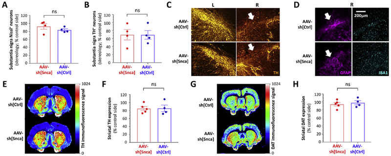
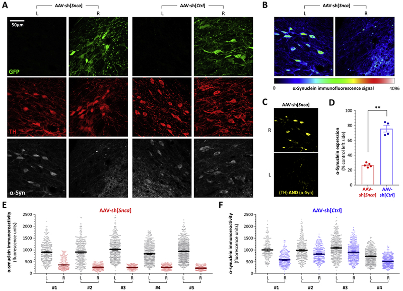
α-Synuclein plays a central role in the pathogenesis of Parkinson's disease (PD); interventions that decrease its expression appear neuroprotective in PD models. Successful translation of these observations into effective therapies will be dependent on the safety of suppressing α-synuclein expression in the adult brain. We investigated long-term α-synuclein knockdown in the adult rat CNS. 8-month old animals received either AAV-sh[Snca] (an RNA interference vector targeting the Snca mRNA transcript) or AAV-sh[Ctrl] (a control vector) unilaterally into the substantia nigra. No signs of systemic toxicity or motor dysfunction were observed in either experimental group over 12 months. Viral transgene expression persisted to 12 months post-inoculation, at which point Snca mRNA expression in substantia nigra dopaminergic neurons of animals that received AAV-sh[Snca] was decreased by ≈90%, and α-synuclein immunoreactivity by >70% relative to the control side. Stereological quantification of Nissl-labeled neurons showed no evidence of neurodegeneration in the substantia nigra 12 months after inoculation with either vector, and we observed abundant dopaminergic neurons with minimal α-synuclein immunoreactivity that appeared otherwise unremarkable in the AAV-sh[Snca] group. Despite the absence of neurodegeneration, some loss of TH expression was evident in nigral neurons after transduction with either vector, presumably a non-specific consequence of vector delivery, cellular transduction, or expression of shRNA or GFP. We conclude that long-term α-synuclein knockdown in the substantia nigra does not cause significant functional deficits in the ascending dopaminergic projection, or neurodegeneration. These findings are encouraging that it may be feasible to target α-synuclein expression therapeutically in PD.


Figures

Figure 1:
Figure 1:. No clinical phenotype in adult rats over the 52 weeks following unilateral inoculation of AAV-sh[Snca] into the substantia nigra
A: Experimental design. All time points are relative to day of intracerebral vector inoculation (day 0). B: Mean ± SE weight (g) of rats over the 52 weeks following inoculation with AAV-sh[Snca] (red, n=5) or AAV-sh[Ctrl] (blue, n=4). Note truncated y-axis. C: The postural instability test was used to evaluate nigrostriatal function. Graphs show mean ± SE displacement necessary to trigger forepaw placement over the 52 weeks following inoculation with AAV-sh[Snca] (red, upper graph, n=5) or AAV-sh[Ctrl] (blue, lower graph, n=4). Note truncated y-axes. For each experimental group, data are superimposed for the negative control right forepaw (ipsilateral to vector inoculation; black triangles) and left forepaw (contralateral to vector inoculation; colored circles). The inset panel shows data redrawn from our prior publication (Zharikov et al., 2015) to illustrate the large unilateral increase in trigger distance that can be detected using this method in the setting of unilateral nigrostriatal dysfunction. In this case the motor deficit was induced by 4 days of rotenone exposure, with the asymmetry arising from neuroprotection following unilateral α-synuclein knockdown (red bars). The dotted lines in the inset graph show the y-axis limits for the main panel. Data analysis in panels B and C was performed by 2-way ANOVA. There were no significant differences between treatment groups (B, C) or sides (C) at any time point.
Figure 2:
Figure 2:. Loss of Snca mRNA expression in the substantia nigra and persistent transgene expression 52 weeks after AAV-sh[Snca] inoculation
A: Photomicrographs of the substantia nigra are shown for animals #2 – #5 in the group receiving AAV-sh[Snca]. Each pair of images shows the substantia nigra on each side (labeled L and R) of the same section. Top row: immunohistochemistry (IHC) for tyrosine hydroxylase (TH; brown) showing the position and integrity of the substantia nigra. Bottom row: RNA in situ hybridization (ISH) for the Snca mRNA transcript (purple). IHC and ISH were performed on adjacent sections. B: Similar to panel A, showing sections from each animal in the group receiving AAV-sh[Ctrl], Top row: IHC for TH (brown). Bottom row: RNA ISH for the Snca mRNA transcript (purple). C: The number of Snca mRNA-expressing cells was counted on each side of the substantia nigra for three sections per animal. Individual data points show the mean value for each animal (AAV-sh[Snca] n=5; AAV-sh[Ctrl] n=4) and side; bars show the group mean ± 95% CI. D: The Snca mRNA ISH signal was quantified by densitometry in the region corresponding to the substantia nigra on each side of three midbrain sections per animal. Individual data points show the mean value for each animal and side; bars show the group mean ± 95% CI. E: Expression of the vector-encoded GFP transgene was determined in the midbrain (left) and forebrain (right) by IHC for GFP (brown), demonstrating persistent presence and expression of the AAV vector 52 weeks post-transduction. The cartoons below the micrographs indicate the planes of the sections shown. Panels C and D were analyzed by 1-way ANOVA with Tukey’s post hoc test. ***p<0.0001, ****p<0.00001.
Figure 3:
Figure 3:. Absence of neurodegeneration but decreased TH expression and astrogliosis in the substantia nigra 52 weeks after inoculation with either AAV-sh[Snca] or AAV-sh[Ctrl]
A: Unbiased stereology was used to quantify Nissl+ neurons on each side of the substantia nigra from rats that received AAV-sh[Snca] (red, left, n=4) or AAV-sh[Ctrl] (blue, right, n=4). Data points show values for individual animals, expressed as % of the control non-transduced side in the same animal. Bars show group mean ± SE. B: Unbiased stereology was used to quantify TH+ neurons on each side of the substantia nigra from rats that received AAV-sh[Snca] (red, left, n=4) or AAV-sh[Ctrl] (blue, right, n=4). Data points show values for individual animals, expressed as % of the control non-transduced side in the same animal. Bars show group mean ± SE. C: Epifluorescence micrographs of the substantia nigra at the point of vector delivery, labeled for TH (orange). Localized loss of TH-expressing cells following delivery of either AAV-sh[Ctrl] (upper row) or AAV-sh[Snca] (lower row) is indicated by white arrows. D: Epifluorescence micrographs of the substantia nigra in adjacent sections to panel C, labeled for GFAP (magenta, left) and IBA-1 (cyan, right). Localized astrogliosis at the site of vector inoculation following delivery of either AAV-sh[Ctrl] (upper row) or AAV-sh[Snca] (lower row) is indicated by white arrows. E – H: Quantitative near-infrared immunofluorescence was employed to quantify TH (E, F) and DAT (G, H) expression in the striatum. E and G show forebrain sections from animals inoculated with AAV-sh[Ctrl] (top) or AAV-sh[Snca] (bottom). The intensity of immunoreactivity is depicted using the color scale shown to the right of each panel. Immunoreactivity for TH (F) and DAT (H) was quantified on each side of the striatum, from rats that received AAV-sh[Snca] (red, left, n=5) or AAV-sh[Ctrl] (blue, right, n=4). Data points show mean values for 5 sections from individual animals expressed as % of the control non-transduced side for the same animal. Bars show group mean ± SE. Data were analyzed by unpaired 2-tailed T-tests. There were no significant differences between vectors.
Figure 4:
Figure 4:. Persistent loss of α-synuclein expression in TH-expressing nigral dopaminergic neurons, 52 weeks after inoculation with AAV-sh[Snca]
A: Confocal planes of the substantia nigra on each side of the same midbrain sections are shown from animals inoculated with AAV-sh[Snca] (left) or AAV-sh[Ctrl] (right). The sections were immunolabeled for GFP (green), TH(red), α-synuclein (white). The scale bar for all 12 images is shown in the top left image. Images from the AAV-sh[Snca] inoculated animal are further analyzed in panels B and C. B: α-Synuclein immunoreactivity is illustrated using a color intensity scale (shown beneath the images) to demonstrate the loss of α-synuclein expression in nigral dopaminergic neurons that received AAV-sh[Snca]. C: A Boolean AND operation was employed to identify pixels from the same confocal plane that showed both TH and α-synuclein immunoreactivity. The resulting image further illustrates loss of α-synuclein expression in dopaminergic neurons 52 weeks after AAV-sh[Snca] inoculation. D, E, F: The TH channel was used to define regions of interest in confocal planes from both sides of multiple midbrain sections, thereby allowing blinded quantification of α-synuclein expression within 350 - 700 dopaminergic neurons from each side of each animal. (E) and (F) show scatter plots of α-synuclein immunoreactivity in individual cells from the control and vector sides of animals inoculated with (E) AAV-sh[Snca] or (F) AAV-sh[Ctrl]; bars show mean and 95% CL (D) shows the mean for each animal expressed as % of the control non-transduced side. Panel D was analyzed using unpaired 2-tailed T-test, **p< 0.01 AAV-sh[Snca] vs AAV-sh[Ctrl].

References

    1. Abeliovich A, et al., 2000. Mice lacking alpha-synuclein display functional deficits in the nigrostriatal dopamine system. Neuron. 25, 239–52. - PubMed
    1. Alarcon-Aris D, et al., 2018. Selective alpha-Synuclein Knockdown in Monoamine Neurons by Intranasal Oligonucleotide Delivery: Potential Therapy for Parkinson’s Disease. Mol Ther. 26, 550–567. - PMC - PubMed
    1. Ansari AM, et al., 2016. Cellular GFP Toxicity and Immunogenicity: Potential Confounders in in Vivo Cell Tracking Experiments. Stem Cell Rev. 12, 553–559. - PMC - PubMed
    1. Benskey MJ, et al., 2018. Silencing Alpha Synuclein in Mature Nigral Neurons Results in Rapid Neuroinflammation and Subsequent Toxicity. Front Mol Neurosci. 11, 36. - PMC - PubMed
    1. Betarbet R, et al., 2000. Chronic systemic pesticide exposure reproduces features of Parkinson’s disease. Nat Neurosci. 3, 1301–6. - PubMed

Publication types