Review
. 2019 Jun:56:78-86.
doi: 10.1016/j.conb.2018.12.003. Epub 2019 Jan 15.

Not all cortical expansions are the same: the coevolution of the neocortex and the dorsal thalamus in mammals

Affiliations
Review

Not all cortical expansions are the same: the coevolution of the neocortex and the dorsal thalamus in mammals

Andrew C Halley et al. Curr Opin Neurobiol. 2019 Jun.

Abstract

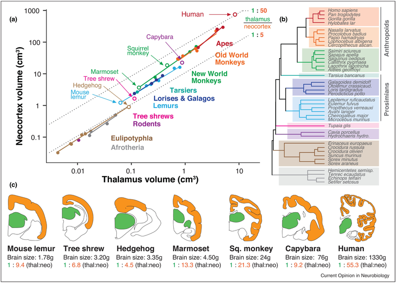
A central question in comparative neurobiology concerns how evolution has produced brains with expanded neocortices, composed of more areas with unique connectivity and functional properties. Some mammalian lineages, such as primates, exhibit exceptionally large cortices relative to the amount of sensory inputs from the dorsal thalamus, and this expansion is associated with a larger number of distinct cortical areas, composing a larger proportion of the cortical sheet. We propose a link between the organization of the neocortex and its expansion relative to the size of the dorsal thalamus, based on a combination of work in comparative neuroanatomy and experimental research.


Conflict of interest statement

Conflicts of Interest

We declare no conflicts of interest.

Figures

Figure 1.
Figure 1.. Primate “neocorticalization” relative to the dorsal thalamus.
Across a wide range of brain sizes, primates exhibit larger neocortices relative to the size of the dorsal thalamus than many mammalian groups. (A) Neocortex volume is plotted against dorsal thalamus volume in log-log coordinates, demonstrating effects of both concerted and mosaic evolution. Concerted evolution is demonstrated by each group having a slope greater than 1 (i.e. more than isometric scaling, or constant proportions across size variation). Mosaic evolution is exemplified by intercept shifts between mammalian lineages. Open circles indicate species shown in coronal section below. (B) A phylogenetic tree of included species, with colors corresponding to mammalian clades in (A). Even after correcting for phylogenetic relatedness, the neocortex always gets larger “faster” than the thalamus does (see text). (C) Coronal sections of selected brains in the dataset above, taken from a similar rostrocaudal level (ventral posterior [VP] nucleus of the thalamus), with associated brain sizes and thalamus:neocortex proportions. Primates, and particularly anthropoids (monkeys and apes) have an unusually large neocortex volume that dwarfs size-associated concerted effects. This mosaic evolution of the primate neocortex is so extreme (over & above size-related concerted effects) that only the largest rodent brain (capybara) has similar thalamus-to-neocortex proportions as the smallest primate brain (mouse lemur). Data from [15,22]. Coronal section outlines (dorsal thalamus in green, neocortex in orange) are from the Comparative Mammalian Brain Collection from the University of Wisconsin, and are not to scale. Thal = dorsal thalamus; neo = neocortex. Marmoset (Callithrix jacchus) thalamus size was estimated from the dataset in [15].
Figure 2.
Figure 2.. Neocortical organization in primates and non-primate mammals with varying brain size.
Maps of cortical organization from flattened sections are drawn from architectonic and functional studies, and are aligned according to log-transformed brain size (vertical axis) with primates to the right, and closely-related euarchontoglire species to the left. Compared with mammals of similar brain size, primates have larger neocortices (parietal, temporal and frontal regions), as well as more non-primary sensory areas. While thalamus size is not available for every species included, larger brains have larger neocortex:thalamus proportions (concerted evolution), and primates have a larger neocortex:thalamus proportions across every brain size than do closely related species (mosaic evolution). All scale bars = 4mm. Maps are redrawn from [61,62,63,64].
Figure 3.
Figure 3.. Neocortex of a rat and mouse lemur drawn to scale.
Although rats and mouse lemurs have a similar size brain (~1.8g), the neocortex of the mouse lemur has a surface area ~3 times that of the rat. The mouse lemur also has a larger number of distinct cortical fields in the expanded area of cortex between S1, V1 and A1. The white region in mouse lemur between area 1/2 and other visual areas (light blue) is likely composed of multiple parietal subdivisions (as in the galago; see Fig 2) that have not been fully characterized. Temporal and frontal cortex have also expanded in mouse lemurs. Modified from [61,62] (rat) and [63] (mouse lemur).
Figure 4.
Figure 4.. Neocortex in normal and bilaterally enucleated opossums and blind mole rats.
Although the neocortex in bilaterally enucleated opossums never receives either spontaneous or sensory driven input from the eyes, an architectonically defined primary visual area (V1 or area 17) is still present. Likewise, the blind mole rat has anophthalmic eyes with skin grown over them, yet they still possess an architectonically defined V1. In blind opossums, what would have been visual cortex is co-opted by the somatosensory and auditory system, and in blind mole rats V1 is activated by auditory inputs. Figures redrawn from [47,49].

Similar articles

Cited by

References

    1. Kaas J: The evolution of neocortex in primates. Prog Br Res 2012, 195:91–102. - PMC - PubMed
    2. Synthetic review of decades of work on primate neocortical organization, with a focus on novel cortical fields observed in primates in comparison to other mammalian lineages.

    1. Goldring A, Krubitzer L: Evolution of parietal cortex in mammals: from manipulation to tool use In Evolution of Nervous Systems, Second Edition, Volume 3 Edited by Krubitzer L, Kaas J. Elsevier; 2017:259–286.
    1. Lui JH, Hansen DV, Kriegstein AR: Development and evolution of the human neocortex. Cell 2011, 146:18–36. - PMC - PubMed
    1. Striedter GF, Srinivasan S, Monuki ES: Cortical folding: when, where, how and why? Annu Rev Neurosci 2015, 38:291–307. - PubMed
    2. A synthetic review of the diverse mechanisms involved in cortical folding in mammalian brain evolution, including a new proposal for the role of radial intercalation in generating tangential expansion.

    1. Mota B, Herculano-Houzel S: Cortical folding scales universally with surface area and thickness, not number of neurons. Science 2015, 349:74–77. - PubMed

Publication types