

Radiological characteristics of myelin oligodendrocyte glycoprotein antibody disease
- PMID: 30658259
- PMCID: PMC6431795
- DOI: 10.1016/j.msard.2019.01.021
Radiological characteristics of myelin oligodendrocyte glycoprotein antibody disease
Abstract
Background: MOG antibody disease is an autoimmune disease of the central nervous system (CNS) characterized by the presence of a serological antibody against myelin oligodendrocyte glycoprotein (MOG). MRI is instrumental in distinguishing neuromyelitis optica spectrum disorder (NMOSD) from multiple sclerosis (MS), but MRI features of MOG disease appear to overlap with NMOSD and MS.
Objectives: In this study we aim to characterize the radiological features of MOG antibody disease and compare the findings with those previously described.
Methods: This is a retrospective study of 26 MOG positive patients. We aim to describe their brain, spinal and orbital MRI features and compare our findings with those previously reported in the literature.
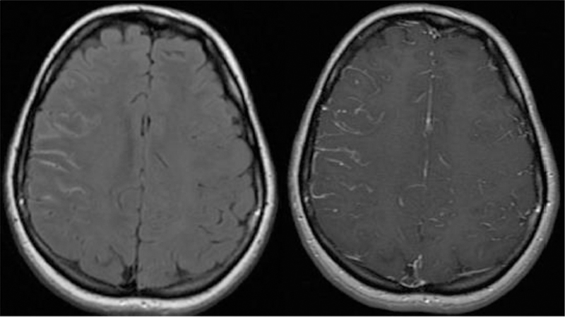
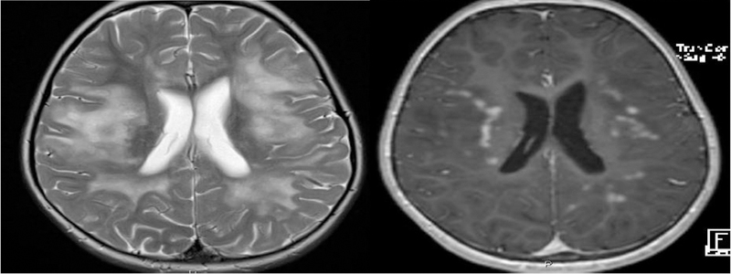
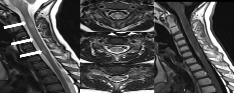
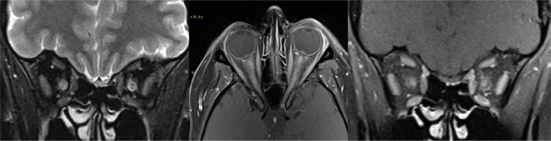
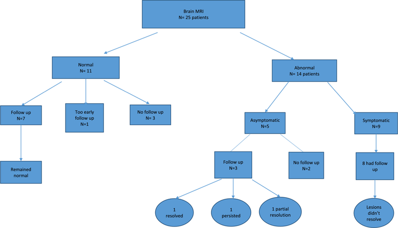
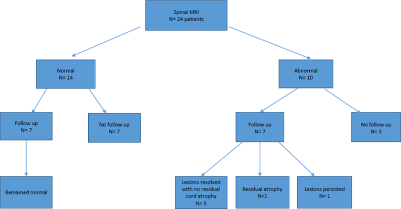
Results: The majority of the abnormal findings was located on orbital MRIs, with more involvement of the anterior structures and bilateral involvement of the optic nerves. Brain abnormalities were distinct from both NMOSD and MS lesions. Spinal cord was the least affected.
Conclusions: This is a dedicated radiological study aiming to characterize the features of MOG antibody disease which might aid in the proper investigation of cases presenting with acquired demyelinating disorders.
Keywords: MOG antibody; Magnetic resonance imaging; NMOSD.
Copyright © 2019 Elsevier B.V. All rights reserved.
Figures

References
-
- Jarius S, Ruprecht K, Kleiter I, Borisow N, Asgari N, Pitarokoili K, et al. MOG-IgG in NMO and related disorders: a multicenter study of 50 patients. Part 1: Frequency, syndrome specificity, influence of disease activity, long-term course, association with AQP4-IgG, and origin. J Neuroinflammation 2016;13(1):279. - PMC - PubMed
-
- Kitley J, Woodhall M, Waters P, Leite MI, Devenney E, Craig J, et al. Myelin-oligodendrocyte glycoprotein antibodies in adults with a neuromyelitis optica phenotype. Neurology 2012;79(12):1273–7. - PubMed
-
- Kitley J, Waters P, Woodhall M, Leite MI, Murchison A, George J, et al. Neuromyelitis optica spectrum disorders with aquaporin-4 and myelin-oligodendrocyte glycoprotein antibodies: a comparative study. JAMA Neurol 2014;71(3):276–83. - PubMed
MeSH terms
Substances
Grants and funding
LinkOut - more resources
Full Text Sources
