. 2019 Jan 17;6(1):8.
doi: 10.3390/bioengineering6010008.

Characterization of Plasma-Derived Extracellular Vesicles Isolated by Different Methods: A Comparison Study

Affiliations

Characterization of Plasma-Derived Extracellular Vesicles Isolated by Different Methods: A Comparison Study

Esther Serrano-Pertierra et al. Bioengineering (Basel). .

Abstract

Extracellular vesicles (EV) are small membrane structures released by cells that act as potent mediators of intercellular communication. The study of EV biology is important, not only to strengthen our knowledge of their physiological roles, but also to better understand their involvement in several diseases. In the field of biomedicine they have been studied as a novel source of biomarkers and drug delivery vehicles. The most commonly used method for EV enrichment in crude pellet involves serial centrifugation and ultracentrifugation. Recently, different protocols and techniques have been developed to isolate EV that imply less time and greater purification. Here we carry out a comparative analysis of three methods to enrich EV from plasma of healthy controls: ultracentrifugation, ExoQuickTM precipitation solution (System Biosciences), and Total Exosome Isolation kit (Invitrogen). Our results show that commercial precipitation reagents are more efficient and enable higher EV enrichment factors compared with traditional ultracentrifugation, although subsequent imaging analysis is not possible with some of them. We hope that this work will contribute to the current research on isolation techniques to assist the progress of clinical applications with diagnostic or therapeutic objectives.

Keywords: enrichment; extracellular vesicles; lateral flow immunoassay; nanoparticle tracking analysis; ultracentrifugation.


Conflict of interest statement

The authors declare that they have no conflict of interest.

Figures

Figure 1
Figure 1
Methods of extracellular vesicles (EV) enrichment. Summary of the main steps of each method for enrichment of plasma-derived EV.
Figure 2
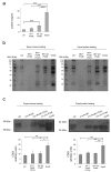
Figure 2
Efficiency of EV enrichment. (a) Protein concentration of the EV fractions isolated was determined by bicinchoninic acid (BCA) assay. The graph shows the mean + SD of the three independent experiments. (b) Coomassie blue staining of the EV fractions. Equal volume and equal amount of protein were loaded for a general protein stain. (c) Representative detection of CD63 in EV fractions by Western blot. Data shown are the mean + SD of three independent experiments. UC: Ultracentrifugation; INV+ProtK: Invitrogen kit and treatment with proteinase K; INV+ProtK (TCA): TCA precipitated protein from INV+ProtK fractions; INV w/o ProtK: Invitrogen kit without digestion with proteinase K; ExoQ: ExoQuick kit. * p < 0.05; ** p < 0.01; *** p < 0.001.
Figure 3
Figure 3
Characterization of plasma-derived EV. Hydrodynamic size distribution profiles of isolated EV measured by (a) DLS and (b) NTA. (c) Mean values + SD of the diameter sizes measured by DLS and NTA (n = 3). (d) Particle concentration of the EV fractions was measured by NTA. The graph shows the mean + SD of three independent experiments. (e) Mean values + SD of the PDI measured by DLS (n = 3). (f) Normalization of EV concentration determined by NTA per protein concentration measured by BCA. The graph shows the mean + SD of three independent experiments. * p < 0.05; ** p < 0.01; *** p < 0.001.
Figure 4
Figure 4
Observation and detection of isolated EV. (a) Transmission electron microscopy images representative of plasma-derived EV by ultracentrifugation or using commercial precipitation reagents (Invitrogen or ExoQuick). (b) Detection of the EV isolated by lateral flow immunoassay (LFIA), using anti-CD9 and anti-CD81 as capture antibodies, and reflectance measurements of AuNPs on each test line (estimated as the peak area of the signal in mV × mm). EV-depleted plasma was used as a negative control (C−). Unbound antiCD63-AuNP captured with anti-IgG were used as system functional verification (Ct).

References

    1. Raposo G., Stoorvogel W. Extracellular vesicles: Exosomes, microvesicles, and friends. J. Cell Biol. 1999;200:373–383. doi: 10.1083/jcb.201211138. - DOI - PMC - PubMed
    1. El Andaloussi S., Mäger I., Breakefield X.O., Wood M.J. Extracellular vesicles: Biology and emerging therapeutic opportunities. Nat. Rev. Drug Discov. 2013;12:347–357. doi: 10.1038/nrd3978. - DOI - PubMed
    1. Cocucci E., Meldolesi J. Ectosomes and exosomes: Shedding the confusion between extracellular vesicles. Trends Cell Biol. 2015;25:364–372. doi: 10.1016/j.tcb.2015.01.004. - DOI - PubMed
    1. Xu R., Greening D.W., Zhu H.J., Takahashi N., Simpson R.J. Extracellular vesicle isolation and characterization: Toward clinical application. J. Clin. Investig. 2016;126:1152–1162. doi: 10.1172/JCI81129. - DOI - PMC - PubMed
    1. Van Dommelen S.M., Vader P., Lakhal S., Kooijman S.A.A., van Solinge W.W., Wood M.J.A., Schiffelers R.M. Microvesicles and exosomes: Opportunities for cell-derived membrane vesicles in drug delivery. J. Control Release. 2012;161:635–644. doi: 10.1016/j.jconrel.2011.11.021. - DOI - PubMed

LinkOut - more resources