Co-Delivery Effect of CD24 on the Immunogenicity and Lethal Challenge Protection of a DNA Vector Expressing Nucleocapsid Protein of Crimean Congo Hemorrhagic Fever Virus
- PMID: 30658445
- PMCID: PMC6356336
- DOI: 10.3390/v11010075
Co-Delivery Effect of CD24 on the Immunogenicity and Lethal Challenge Protection of a DNA Vector Expressing Nucleocapsid Protein of Crimean Congo Hemorrhagic Fever Virus
Abstract
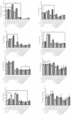
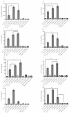
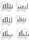
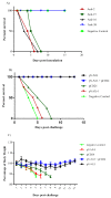
Crimean Congo hemorrhagic fever virus (CCHFV) is the causative agent of a globally-spread tick-borne zoonotic infection, with an eminent risk of fatal human disease. The imminent public health threat posed by the disseminated virus activity and lack of an approved therapeutic make CCHFV an urgent target for vaccine development. We described the construction of a DNA vector expressing a nucleocapsid protein (N) of CCHFV (pV-N13), and investigated its potential to stimulate the cytokine and total/specific antibody responses in BALB/c and a challenge experiment in IFNAR-/- mice. Because of a lack of sufficient antibody stimulation towards the N protein, we have selected cluster of differentiation 24 (CD24) protein as a potential adjuvant, which has a proliferative effect on B and T cells. Overall, our N expressing construct, when administered solely or in combination with the pCD24 vector, elicited significant cellular and humoral responses in BALB/c, despite variations in the particular cytokines and total antibodies. However, the stimulated antibodies produced as a result of the N protein expression have shown no neutralizing ability in the virus neutralization assay. Furthermore, the challenge experiments revealed the protection potential of the N expressing construct in an IFNAR -/- mice model. The cytokine analysis in the IFNAR-/- mice showed an elevation in the IL-6 and TNF-alpha levels. In conclusion, we have shown that targeting the S segment of CCHFV can be considered for a practical way to develop a vaccine against this virus, because of its ability to induce an immune response, which leads to protection in the challenge assays in the interferon (IFN)-gamma defective mice models. Moreover, CD24 has a prominent immunologic effect when it co-delivers with a suitable foreign gene expressing vector.
Keywords: CCHFV; CD24; Cytokines; IFNAR−/− mice; Lethal Challenge; nucleocapsid.
Conflict of interest statement
The authors declare no conflict of interest.
Figures


Similar articles
-
Bovine Herpesvirus Type 4 (BoHV-4) Vector Delivering Nucleocapsid Protein of Crimean-Congo Hemorrhagic Fever Virus Induces Comparable Protective Immunity against Lethal Challenge in IFNα/β/γR-/- Mice Models.Viruses. 2019 Mar 9;11(3):237. doi: 10.3390/v11030237. Viruses. 2019. PMID: 30857305 Free PMC article.
-
Immunization with DNA Plasmids Coding for Crimean-Congo Hemorrhagic Fever Virus Capsid and Envelope Proteins and/or Virus-Like Particles Induces Protection and Survival in Challenged Mice.J Virol. 2017 Apr 28;91(10):e02076-16. doi: 10.1128/JVI.02076-16. Print 2017 May 15. J Virol. 2017. PMID: 28250124 Free PMC article.
-
A DNA vaccine for Crimean-Congo hemorrhagic fever protects against disease and death in two lethal mouse models.PLoS Negl Trop Dis. 2017 Sep 18;11(9):e0005908. doi: 10.1371/journal.pntd.0005908. eCollection 2017 Sep. PLoS Negl Trop Dis. 2017. PMID: 28922426 Free PMC article.
-
The Role of Nucleocapsid Protein (NP) in the Immunology of Crimean-Congo Hemorrhagic Fever Virus (CCHFV).Viruses. 2024 Sep 30;16(10):1547. doi: 10.3390/v16101547. Viruses. 2024. PMID: 39459881 Free PMC article. Review.
-
Recent advances in understanding Crimean-Congo hemorrhagic fever virus.F1000Res. 2018 Oct 29;7:F1000 Faculty Rev-1715. doi: 10.12688/f1000research.16189.1. eCollection 2018. F1000Res. 2018. PMID: 30416710 Free PMC article. Review.
Cited by
-
Crimean-Congo Hemorrhagic Fever Virus: Progress in Vaccine Development.Diagnostics (Basel). 2023 Aug 19;13(16):2708. doi: 10.3390/diagnostics13162708. Diagnostics (Basel). 2023. PMID: 37627967 Free PMC article. Review.
-
Crimean-Congo Hemorrhagic Fever Mouse Model Recapitulating Human Convalescence.J Virol. 2019 Aug 28;93(18):e00554-19. doi: 10.1128/JVI.00554-19. Print 2019 Sep 15. J Virol. 2019. PMID: 31292241 Free PMC article.
-
GP38 as a vaccine target for Crimean-Congo hemorrhagic fever virus.NPJ Vaccines. 2023 May 20;8(1):73. doi: 10.1038/s41541-023-00663-5. NPJ Vaccines. 2023. PMID: 37210392 Free PMC article.
-
Immunogenicity of a DNA-Based Sindbis Replicon Expressing Crimean-Congo Hemorrhagic Fever Virus Nucleoprotein.Vaccines (Basel). 2021 Dec 16;9(12):1491. doi: 10.3390/vaccines9121491. Vaccines (Basel). 2021. PMID: 34960237 Free PMC article.
-
Construction and evaluation of DNA vaccine encoding Crimean Congo hemorrhagic fever virus nucleocapsid protein, glycoprotein N-terminal and C-terminal fused with LAMP1.Front Cell Infect Microbiol. 2023 Mar 21;13:1121163. doi: 10.3389/fcimb.2023.1121163. eCollection 2023. Front Cell Infect Microbiol. 2023. PMID: 37026060 Free PMC article.
References
-
- Garrison A.R., Shoemaker C.J., Golden J.W., Fitzpatrick C.J., Suschak J.J., Richards M.J., Badger C.V., Six C.M., Martin J.D., Hannaman D., et al. A DNA vaccine for Crimean-Congo hemorrhagic fever protects against disease and death in two lethal mouse models. PLoS Negl. Trop. Dis. 2017;11:e0005908. doi: 10.1371/journal.pntd.0005908. - DOI - PMC - PubMed
Publication types
MeSH terms
Substances
Grants and funding
LinkOut - more resources
Full Text Sources
Other Literature Sources
Molecular Biology Databases

