. 2019 Jan 17;8(1):27.
doi: 10.3390/antiox8010027.

KRIT1 Loss-Of-Function Associated with Cerebral Cavernous Malformation Disease Leads to Enhanced S-Glutathionylation of Distinct Structural and Regulatory Proteins

Affiliations

KRIT1 Loss-Of-Function Associated with Cerebral Cavernous Malformation Disease Leads to Enhanced S-Glutathionylation of Distinct Structural and Regulatory Proteins

Laura Cianfruglia et al. Antioxidants (Basel). .

Abstract

Loss-of-function mutations in the KRIT1 gene are associated with the pathogenesis of cerebral cavernous malformations (CCMs), a major cerebrovascular disease still awaiting therapies. Accumulating evidence demonstrates that KRIT1 plays an important role in major redox-sensitive mechanisms, including transcriptional pathways and autophagy, which play major roles in cellular homeostasis and defense against oxidative stress, raising the possibility that KRIT1 loss has pleiotropic effects on multiple redox-sensitive systems. Using previously established cellular models, we found that KRIT1 loss-of-function affects the glutathione (GSH) redox system, causing a significant decrease in total GSH levels and increase in oxidized glutathione disulfide (GSSG), with a consequent deficit in the GSH/GSSG redox ratio and GSH-mediated antioxidant capacity. Redox proteomic analyses showed that these effects are associated with increased S-glutathionylation of distinct proteins involved in adaptive responses to oxidative stress, including redox-sensitive chaperonins, metabolic enzymes, and cytoskeletal proteins, suggesting a novel molecular signature of KRIT1 loss-of-function. Besides providing further insights into the emerging pleiotropic functions of KRIT1, these findings point definitively to KRIT1 as a major player in redox biology, shedding new light on the mechanistic relationship between KRIT1 loss-of-function and enhanced cell sensitivity to oxidative stress, which may eventually lead to cellular dysfunctions and CCM disease pathogenesis.

Keywords: KRIT1; altered redox homeostasis and signaling; cerebral cavernous malformations; mass spectrometry; oxidative post-translational modifications; oxidative stress; protein S-glutathionylation; redox proteomics.


Conflict of interest statement

The authors declare no conflicts of interest.

Figures

Figure 1
Figure 1
KRIT1 loss-of-function induces GSH depletion. (A,B) The amounts of total glutathione (GSH+GSSG in GSH equivalents) (A) and oxidized glutathione (GSSG) (B) were quantified in K−/− cells left untreated or treated for 4 h with liposome-encapsulated GSH (5 mM final concentration) by an established enzymatic recycling assay, as described in Materials and Methods. K9/6 cells were used as control. (C) Histogram representing the GSH/GSSG ratio. (D) Total antioxidant capacity for peroxyl radicals in K9/6 and K−/− cells, as determined by the total oxyradical scavenging capacity (TOSC) assay described in Materials and Methods. For all measurements, TOSC values were referred to protein concentration counterparts. Results are reported as mean values ± standard deviation (S.D.) of six different experiments. Asterisks above histogram bars indicate significant differences (p ≤ 0.05) between groups of means (post hoc comparison).
Figure 2
Figure 2
GSH delivery rescues the cellular redox imbalance induced by KRIT1 loss-of-function. (A) Measurement of ROS levels in K−/− cells left untreated or treated for 4 h with 5 mM GSH delivered by liposomes. K9/6 cells were used as control. The cell-permeant carboxy-H2DCFDA (C400) probe was used for ROS labeling and measurements were performed by flow cytometry, as described in Materials and Methods. ROS concentrations are reported as fluorescence arbitrary units normalized to the initial mean control values, and represent the mean ± S.D. of three independent experiments. Asterisk indicates significant differences (p < 0.0001). (B) Flow cytometric graph representative of the data reported in (A). Gray line = K9/6 cells; red line = K−/− cells; blue line = K−/− cells plus GSH.
Figure 3
Figure 3
KRIT1 loss-of-function affects the activity of GSH-dependent antioxidant enzymes. Glutathione-S-transferase (GST) (A), glutathione peroxidase (GPX) (B), glutathione reductase (GR) (C), and catalase (CAT) activity (D) in K9/6 and K−/− cells left untreated or treated for 4 h with 5 mM GSH encapsulated within liposomes was measured by specific enzymatic assays, as described in Materials and Methods. Results are reported as mean values ± S.D. of six different experiments. Asterisks above histogram bars indicate significant differences (p ≤ 0.05) between groups of means (post hoc comparison).
Figure 4
Figure 4
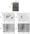
KRIT1 loss-of-function induces changes in protein S-glutathionylation pattern. Total protein extracts of K−/− cells left untreated or treated for 4 h with 5 mM GSH encapsulated within liposomes were separated by monodimensional (A) and two-dimensional electrophoresis (BE) under nonreducing conditions, and analyzed by immunoblotting with an anti-GSH antibody to detect protein S-glutathionylation adducts (A–C), or by colloidal Coomassie staining to identify protein spots by mass spectrometry analysis (D,E). K9/6 cells were used as control. (A) Equal amounts of protein extracts (30 μg) of the indicated cells were analyzed by SDS-PAGE and Western blotting under nonreducing conditions. (B–E) Equal amounts of protein extracts (200 μg) of K−/− (B,D) and K9/6 (C,E) cells were analyzed by two-dimensional (2-D) electrophoresis and Western blotting. Parallel experiments were performed for Western blotting (B,C) and colloidal Coomassie staining (D,E) analyses. Digitalized images of PVDF membranes and colloidal Coomassie-stained gels were acquired and analyzed with a dedicated software that ensured spot matching and relative quantitation. Gel spots corresponding to immunoreactive signals were manually excised from 2D-gels and further subjected to mass spectrometric analysis. Experiments were performed in technical duplicate on two biological replicates.
Figure 5
Figure 5
Identification of S-glutathionylated cysteine 328 in vimentin. Collision induced dissociation fragmentation spectrum of the doubly charged (m/z 862.3) modified peptide (322–334) from vimentin-bearing S-glutathionylated Cys328. This peptide was isolated in spot 10 from K−/− cells (Figure 4B,E). Assigned fragments are reported in the spectrum, together with the peptide sequence where the modified residue is underlined. Glu, glutathione.
Figure 6
Figure 6
Confirmation of HSP60 S-glutathionylation induced by KRIT1 loss-of-function. (A) Lysates from K9/6 and K−/− cells were immunoprecipitated with protein G agarose-coupled anti-HSP60 (IP: Hsp60) and subjected to Western blotting (WB) with anti-GSH antibody. Blots were then stripped and reprobed with anti-HSP60 antibody to ensure equal immunoprecipitation of HSP60 proteins. Mouse IgG was used as negative control for immunoprecipitation. (B) The histogram indicates mean (±S.E., n = 3) levels of GSH-Hsp60 relative to total Hsp60 in IP samples, as quantified by densitometric analysis of WB bands. Normalized optical density values were expressed as relative protein level units. Asterisks indicate significant differences versus K9/6 cells (p ≤ 0.01).
Figure 7
Figure 7
Redox-dependent changes in PSSG occur in human brain microvascular endothelial cells upon KRIT1 knockdown. Human brain microvascular endothelial cells (hBMEC) grown under standard conditions were transfected with either KRIT1-targeting siRNA (si-KRIT1) or a scrambled control (si-CTR). Cells were then either left untreated or treated with Tiron, lysed, and analyzed by Western blotting under nonreducing conditions with an anti-GSH antibody to detect protein S-glutathionylation adducts, and then compared with KRIT1 protein expression levels. β-actin was used as internal loading control for Western blot normalization. Results are representative of three separate experiments. Notice that KRIT1 knockdown in human brain microvascular endothelial cells leads to a significant increase in the levels of protein S-glutathionylation adducts, which are significantly reverted by cell treatment with the ROS scavenger Tiron.
Figure 8
Figure 8
Increased levels of S-glutathionylated proteins occur in endothelial cells lining human CCM lesions. Immunohistochemical (IHC) analysis of S-glutathionylated proteins in histological sections of a representative human CCM surgical specimen containing a cluster of CCM vessels (A) and perilesional normal vessels serving as internal negative controls (B). Scale bar: 200 µm. Notice that a significant positive IHC staining for S-glutathionylated proteins is evident in endothelial cells lining the lumen of CCM lesions (A, arrows), as compared with endothelial cells lining the lumen of perilesional normal vessels (B, asterisks).
Figure 9
Figure 9
Schematic model representing the S-glutathionylation of distinct structural and regulatory proteins as a novel molecular signature of KRIT1 loss-of-function. The altered intracellular redox homeostasis caused by KRIT1 loss-of-function affects the glutathione (GSH) redox system, leading to a significant decrease in total GSH levels and increase in oxidized glutathione disulfide (GSSG), with a consequent deficit in the GSH/GSSG redox ratio and GSH-mediated antioxidant capacity. These effects are associated with increased S-glutathionylation of distinct proteins involved in adaptive responses to oxidative stress, including redox-sensitive chaperonins, metabolic enzymes, and cytoskeletal proteins, suggesting a novel molecular signature of KRIT1 loss-of-function that could contribute to its emerging pleiotropic effects in the pathogenesis of CCM disease.

Similar articles

Cited by

References

    1. Batra S., Lin D., Recinos P.F., Zhang J., Rigamonti D. Cavernous malformations: Natural history, diagnosis and treatment. Nat. Rev. Neurol. 2009;5:659–670. doi: 10.1038/nrneurol.2009.177. - DOI - PubMed
    1. Choquet H., Pawlikowska L., Lawton M.T., Kim H. Genetics of cerebral cavernous malformations: Current status and future prospects. J. Neurosurg. Sci. 2015;59:211–220. - PMC - PubMed
    1. Retta S.F., Glading A.J. Oxidative stress and inflammation in cerebral cavernous malformation disease pathogenesis: Two sides of the same coin. Int. J. Biochem. Cell Biol. 2016;81:254–270. doi: 10.1016/j.biocel.2016.09.011. - DOI - PMC - PubMed
    1. Trapani E., Retta S.F. Cerebral cavernous malformation (CCM) disease: From monogenic forms to genetic susceptibility factors. J. Neurosurg. Sci. 2015;59:201–209. - PubMed
    1. Fontanella M., Bacigaluppi S. Treatment of cerebral cavernous malformations: Where do we stand. J. Neurosurg. Sci. 2015;59:199–200. - PubMed