

Cancer-Associated Intermediate Conductance Ca2+-Activated K⁺ Channel KCa3.1
- PMID: 30658505
- PMCID: PMC6357066
- DOI: 10.3390/cancers11010109
Cancer-Associated Intermediate Conductance Ca2+-Activated K⁺ Channel KCa3.1
Abstract
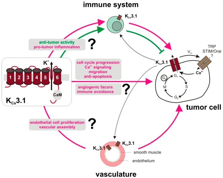
Several tumor entities have been reported to overexpress KCa3.1 potassium channels due to epigenetic, transcriptional, or post-translational modifications. By modulating membrane potential, cell volume, or Ca2+ signaling, KCa3.1 has been proposed to exert pivotal oncogenic functions in tumorigenesis, malignant progression, metastasis, and therapy resistance. Moreover, KCa3.1 is expressed by tumor-promoting stroma cells such as fibroblasts and the tumor vasculature suggesting a role of KCa3.1 in the adaptation of the tumor microenvironment. Combined, this features KCa3.1 as a candidate target for innovative anti-cancer therapy. However, immune cells also express KCa3.1 thereby contributing to T cell activation. Thus, any strategy targeting KCa3.1 in anti-cancer therapy may also modulate anti-tumor immune activity and/or immunosuppression. The present review article highlights the potential of KCa3.1 as an anti-tumor target providing an overview of the current knowledge on its function in tumor pathogenesis with emphasis on vasculo- and angiogenesis as well as anti-cancer immune responses.
Keywords: (1-[(2-chlorophenyl) diphenylmethyl]-pyrazole; 1-EBIO; 1-Ethyl-1,3-dihydro-2H-benzimidazol-2-one; 17-estradiol; BK; E2; KCa3.1; TRAM-34; big conductance Ca2+- and voltage-activated K+ channels; intermediate conductance calcium-activated K+ channel.
Conflict of interest statement
S.M.H has a research collaboration with Novocure, Haifa, Israel.
Figures
Similar articles
-
Intermediate-conductance calcium-activated potassium channel KCa3.1 and chloride channel modulate chemokine ligand (CCL19/CCL21)-induced migration of dendritic cells.Transl Res. 2015 Jul;166(1):89-102. doi: 10.1016/j.trsl.2014.11.010. Epub 2014 Dec 20. Transl Res. 2015. PMID: 25583444 Free PMC article.
-
Functional Roles of the Ca2+-activated K+ Channel, KCa3.1, in Brain Tumors.Curr Neuropharmacol. 2018;16(5):636-643. doi: 10.2174/0929867324666170713103621. Curr Neuropharmacol. 2018. PMID: 28707595 Free PMC article. Review.
-
Effects of Intermediate-Conductance Ca(2+)-Activated K(+) Channels on Human Endometrial Carcinoma Cells.Cell Biochem Biophys. 2015 Jun;72(2):515-25. doi: 10.1007/s12013-014-0497-0. Cell Biochem Biophys. 2015. PMID: 25608633
-
Inhibitory actions by ibandronate sodium, a nitrogen-containing bisphosphonate, on calcium-activated potassium channels in Madin-Darby canine kidney cells.Toxicol Rep. 2015 Aug 28;2:1182-1193. doi: 10.1016/j.toxrep.2015.08.010. eCollection 2015. Toxicol Rep. 2015. PMID: 28962460 Free PMC article.
-
Ca2+-Activated K+ Channel KCa3.1 as a Therapeutic Target for Immune Disorders.Biol Pharm Bull. 2018;41(8):1158-1163. doi: 10.1248/bpb.b18-00078. Biol Pharm Bull. 2018. PMID: 30068864 Review.
Cited by
-
pH-Channeling in Cancer: How pH-Dependence of Cation Channels Shapes Cancer Pathophysiology.Cancers (Basel). 2020 Sep 2;12(9):2484. doi: 10.3390/cancers12092484. Cancers (Basel). 2020. PMID: 32887220 Free PMC article. Review.
-
Depressive effectiveness of vigabatrin (γ-vinyl-GABA), an antiepileptic drug, in intermediate-conductance calcium-activated potassium channels in human glioma cells.BMC Pharmacol Toxicol. 2021 Jan 13;22(1):6. doi: 10.1186/s40360-021-00472-3. BMC Pharmacol Toxicol. 2021. PMID: 33441172 Free PMC article.
-
The N and C-termini of SPCA2 regulate differently Kv10.1 function: role in the collagen 1-induced breast cancer cell survival.Am J Cancer Res. 2021 Jan 1;11(1):251-263. eCollection 2021. Am J Cancer Res. 2021. PMID: 33520372 Free PMC article.
-
Effects of KCa channels on biological behavior of trophoblasts.Open Life Sci. 2022 Sep 3;17(1):1043-1052. doi: 10.1515/biol-2022-0462. eCollection 2022. Open Life Sci. 2022. PMID: 36118166 Free PMC article.
-
Imaging of KCa 3.1 Channels in Tumor Cells with PET and Small-Molecule Fluorescent Probes.ChemMedChem. 2023 Jan 17;18(2):e202200551. doi: 10.1002/cmdc.202200551. Epub 2022 Nov 22. ChemMedChem. 2023. PMID: 36315933 Free PMC article.
References
-
- Fanger C.M., Ghanshani S., Logsdon N.J., Rauer H., Kalman K., Zhou J., Beckingham K., Chandy K.G., Cahalan M.D., Aiyar J. Calmodulin mediates calcium-dependent activation of the intermediate conductance KCa channel, IKCa1. J. Biol. Chem. 1999;274:5746–5754. doi: 10.1074/jbc.274.9.5746. - DOI - PubMed
Publication types
LinkOut - more resources
Full Text Sources
Other Literature Sources
Miscellaneous
