

Extracellular vesicles in bone: "dogrobbers" in the "eternal battle field"
- PMID: 30658653
- PMCID: PMC6339294
- DOI: 10.1186/s12964-019-0319-5
Extracellular vesicles in bone: "dogrobbers" in the "eternal battle field"
Abstract
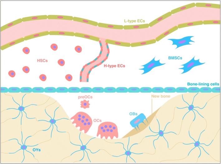
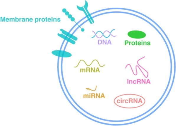
Throughout human life, bone is constantly in a delicate dynamic equilibrium of synthesis and resorption, hosting finely-tuned bone mineral metabolic processes for bone homeostasis by collaboration or symphony among several cell types including osteoclasts (OCs), osteoblasts (OBs), osteocytes (OYs), vascular endothelial cells (ECs) and their precursors. Beyond these connections, a substantial level of communication seems to occur between bone and other tissues, and together, they form an organic unit linked to human health and disease. However, the current hypothesis, which includes growth factors, hormones and specific protein secretion, incompletely explains the close connections among bone cells or between bone and other tissues. Extracellular vesicles (EVs) are widely-distributed membrane structures consisting of lipid bilayers, membrane proteins and intravesicular cargo (including proteins and nucleic acids), ranging from 30 nm to 1000 nm in diameter, and their characters have been highly conserved throughout evolution. EVs have targeting abilities and the potential to transmit multidimensional, abundant and complicated information, as powerful and substantial "dogrobbers" mediating intercellular communications. As research has progressed, EVs have gradually become thought of as "dogrobbers" in bone tissue-the "eternal battle field" -in a delicate dynamic balance of destruction and reconstruction. In the current review, we give a brief description of the major constituent cells in bone tissues and explore the progress of current research on bone-derived EVs. In addition, this review also discusses in depth not only potential directions for future research to breakthrough in this area but also problems existing in current research that need to be solved for a better understanding of bone tissues.
Keywords: Bone; Exosomes; Extracellular vesicles; Intercellular communications; Signalling pathways.
Conflict of interest statement
Consent for publication
Not applicable.
Competing interests
The authors declared that they no competing interests.
Publisher’s Note
Springer Nature remains neutral with regard to jurisdictional claims in published maps and institutional affiliations.
Figures
References
-
- Cappariello A, Ponzetti M, Rucci N. The “soft” side of the bone: unveiling its endocrine functions. Horm Mol Biol Clin Investig. 2016;28:5–20. - PubMed
-
- Fernandez-Real JM, Izquierdo M, Ortega F, Gorostiaga E, Gomez-Ambrosi J, Moreno-Navarrete JM, Fruhbeck G, Martinez C, Idoate F, Salvador J, et al. The relationship of serum osteocalcin concentration to insulin secretion, sensitivity, and disposal with hypocaloric diet and resistance training. J Clin Endocrinol Metab. 2009;94:237–245. doi: 10.1210/jc.2008-0270. - DOI - PubMed
Publication types
MeSH terms
LinkOut - more resources
Full Text Sources
Research Materials
