. 2019 Apr 1;79(7):1646-1657.
doi: 10.1158/0008-5472.CAN-18-1602. Epub 2019 Jan 18.

MNK1/NODAL Signaling Promotes Invasive Progression of Breast Ductal Carcinoma In Situ

Affiliations

MNK1/NODAL Signaling Promotes Invasive Progression of Breast Ductal Carcinoma In Situ

Qianyu Guo et al. Cancer Res. .

Erratum in

  • Correction: MNK1/NODAL Signaling Promotes Invasive Progression of Breast Ductal Carcinoma In Situ.
    Guo Q, Li VZ, Nichol JN, Huang F, Yang W, Preston SEJ, Talat Z, Lefrère H, Yu H, Zhang G, Basik M, Gonçalves C, Zhan Y, Plourde D, Su J, Torres J, Marques M, Habyan SA, Bijian K, Amant F, Wichter M, Behbod F, McCaffrey L, Alaoui-Jamali M, Giannakopoulos NV, Brackstone M, Postovit LM, Del Rincón SV, Miller WH Jr. Guo Q, et al. Cancer Res. 2024 Apr 15;84(8):1373. doi: 10.1158/0008-5472.CAN-24-0461. Cancer Res. 2024. PMID: 38616659 No abstract available.

Abstract

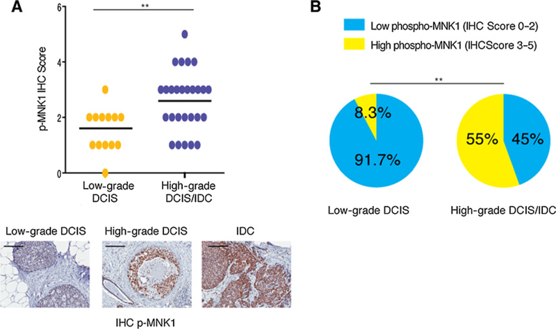
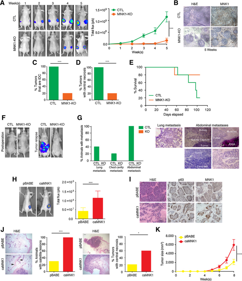
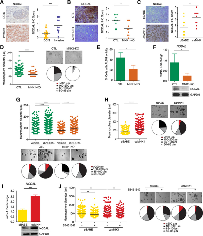
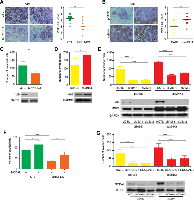
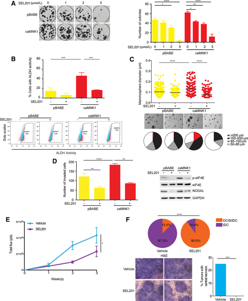
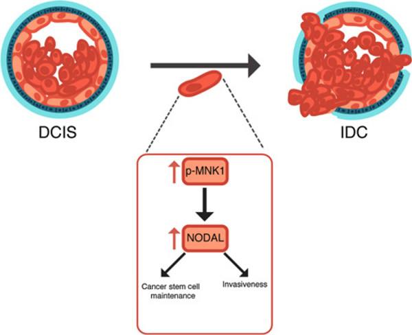
The mechanisms by which breast cancers progress from relatively indolent ductal carcinoma in situ (DCIS) to invasive ductal carcinoma (IDC) are not well understood. However, this process is critical to the acquisition of metastatic potential. MAPK-interacting serine/threonine-protein kinase 1 (MNK1) signaling can promote cell invasion. NODAL, a morphogen essential for embryogenic patterning, is often reexpressed in breast cancer. Here we describe a MNK1/NODAL signaling axis that promotes DCIS progression to IDC. We generated MNK1 knockout (KO) or constitutively active MNK1 (caMNK1)-expressing human MCF-10A-derived DCIS cell lines, which were orthotopically injected into the mammary glands of mice. Loss of MNK1 repressed NODAL expression, inhibited DCIS to IDC conversion, and decreased tumor relapse and metastasis. Conversely, caMNK1 induced NODAL expression and promoted IDC. The MNK1/NODAL axis promoted cancer stem cell properties and invasion in vitro. The MNK1/2 inhibitor SEL201 blocked DCIS progression to invasive disease in vivo. In clinical samples, IDC and DCIS with microinvasion expressed higher levels of phospho-MNK1 and NODAL versus low-grade (invasion-free) DCIS. Cumulatively, our data support further development of MNK1 inhibitors as therapeutics for preventing invasive disease. SIGNIFICANCE: These findings provide new mechanistic insight into progression of ductal carcinoma and support clinical application of MNK1 inhibitors to delay progression of indolent ductal carcinoma in situ to invasive ductal carcinoma.


Conflict of interest statement

Disclosure of Potential Conflicts of Interest

No potential conflicts of interest were disclosed.

Figures

Figure 1.
Figure 1.
Increased MNK1 activity in IDC and high-grade DCIS. A, Phospho-MNK1 levels are higher in high-grade DCIS/IDC samples than low-grade DCIS samples. Scale bar,200 μm. B, Increased percentage of samples with high phospho-MNK1 staining in high-grade DCIS/IDC compared with low-grade DCIS. _**, P < 0.01.
Figure 2.
Figure 2.
MNK1 regulates the DCIS-IDC transition in vivo. A, DCIS-Luc CTL and MNK1-KO tumor outgrowth is measured by IVIS imaging. B, MNK1 knockout is retained in the DCIS-Luc MNK1-KO xenografts as confirmed by IHC. Scale bar, 50 μm. C, All DCIS-Luc CTL xenografts have progressed into IDC, while only 20% DCIS-Luc MNK1-KO tumors have progressed to an IDC-like morphology. D, All DCIS-Luc CTL xenografts have central necrosis, while only 20% DCIS-Luc MNK1-KO tumors have central necrosis. E, Survival curve of mice receiving DCIS-Luc CTL/MNK1-KO cells. F, Representative IVIS imaging showing complete tumor removal postoperation and tumor recurrence in animals receiving DCIS-Luc CTL cells. G, Percentage of animals presented with metastasis at different sites and representative images of metastasis in various tissues of mice receiving DCIS-Luc CTL/MNK1-KO cells. Scale bar, 200 μm. H, Tumor outgrowth is measured by IVIS imaging. I, DCIS-Luc pBABE xenografts maintain DCIS morphology, while DCIS-Luc caMNK1 tumors have progressed into a mixed morphology of DCIS/IDC. caMNKI overexpression is maintained in the xenografts as confirmed by IHC. Scale bar, 50 μm. J, 100% of mice with DCIS-Luc caMNK1 tumors have micrometastasis in the mammary gland, while 30% of DCIS-Luc pBABE have micrometastasis. Arrows, micrometastases. Sixty percent DCIS-Luc caMNK1 and 20% DCIS-Luc pBABE tumors have central necrosis. Scale bar, 200 μm. K, DCIS-Luc caMNK1 xenografts present with growth advantage over DCIS-Luc pBABE controls. H&E, hematoxylin and eosin. ***, P < 0.001; ****, P< 0.0001.
Figure 3.
Figure 3.
MNK1 expression regulates NODAL morphogen expression. A, NODAL expression in DCIS versus invasive human breast samples. Scale bar, 200 μm. B, DCIS-Luc MNK1-KO xenografts have decreased NODAL levels compared with DCIS-Luc CTL tumors. Representative images are shown. Scale bar, 200 μm. C, DCIS-Luc caMNK1 xenografts have increased NODAL levels compared with DCIS-Luc pBABE tumors. Representative images are shown. Scale bar, 200 μm. D, DCIS-Luc MNK1-KO cells have mammosphere size in low adherent culture. Scale bar, 200 μm E, DCIS-Luc MNK1-KO cells have reduced ALDH+ populations. F, DCIS-Luc MNK1-KO mammospheres express lower NODAL mRNA levels. G, rhNODAL treatment increases mammosphere size in DCIS-Luc CTL/MNK1-KO cells. Scale bar, 200 μm. H, DCIS-Luc caMNK1 overexpression increases mammosphere size in low adherent culture. Scale bar, 200 μm. I, DCIS-Luc caMNK1 mammospheres express higher NODAL mRNA levels. J, DCIS-Luc pBABE/caMNK1 mammosphere sizes can both be reduced by 10 μmol/LSB431542, a NODAL pathway inhibitor. Scale bar, 200 μm. *, P < 0.05;**, P< 0.01; ***, P< 0.001; ****, P < 0.0001.
Figure 4.
Figure 4.
The MNK1/NODAL axis regulates migration and invasion. A, DCIS-Luc MNK-KO xenografts have decreased VIMENTIN (VIM) levels compared with DCIS-Luc CTL tumors. Representative images from two tumors are shown. Scale bar, 200 μm. B, DCIS-Luc caMNK1 xenografts have increased VIMENTIN (VIM) levels compared with DCIS-Luc pBABE control tumors. Representative images from two tumors are shown. Scale bar, 200 μm. C, DCIS-Luc MNK1-KO cells have impaired capacity to migrate and to invade through Collagen I in transwells. D, DCIS-LuccaMNK1 cells showed increased capacity to migrate and invade through Collagen I in transwells. E, VIMENTIN knockdown by siRNA decreases the invasive capacity of DCIS-Luc pBABE/caMNK1 cells. F, rhNODAL treatment increases the migration and invasion of DCIS-Luc MNK1-KO cells. G, Transient knockdown of NODAL decreases the invasive capacity of DCIS-Luc pBABE/caMNK1 cells. *, P< 0.05;**, P < 0.01; ****, P < 0.0001.
Figure 5.
Figure 5.
Pharmacologically targeting MNK1 inhibits the DCIS/IDC transition A. SEL201 inhibits DCIS-Luc pBABE/caMNK1 colony formation in a dose-dependent manner. B, Two μmol/L SEL201 decreases the ALDH+ population in DCIS-Luc caMNK1 cells. C, Five μmol/L SEL201 decreases mammosphere size formed by DCIS-Luc caMNK1 cells. Scale bar, 200 μm D, Two μmol/L SEL201 decreases the invasive capacity of DCIS-Luc caMNK1 cells. E, Primary tumor outgrowth over 3 weeks in vehicle versus SEL201-treated animals injected with DCIS-Luc CTL cells. F, MNK inhibitor SEL201 treatment slows down DCIS to IDC progression in nude mice injected with DCIS-Luc CTL cells and decreases the percentage of tumors with detectable central necrosis. *, P < 0.05; **, P < 0.01; ***, P < 0.001; ****, P < 0.0001.
Figure 6.
Figure 6.
A model depicting the MNK1/NODAL axis during the DCIS to IDC transition. Our data suggest that high MNK1 activity promotes the expression of NODAL. NODAL, in turn, promotes cell invasion and cancer stem cell maintenance.

References

    1. Martinez-Perez C, Turnbull AK, Ekatah GE, Arthur LM, Sims AH, Thomas JS, et al. Current treatment trends and the need for better predictive tools in the management of ductal carcinoma in situ of the breast. Cancer Treat Rev 2017;55:163–72. - PubMed
    1. Wang X, Flynn A, Waskiewicz AJ, Webb BL, Vries RG, Baines IA, et al. The phosphorylation of eukaryotic initiation factor eIF4E in response to phorbol esters, cell stresses, and cytokines is mediated by distinct MAP kinase pathways. J Biol Chem 1998; 273:9373–7. - PubMed
    1. Bhat M, Robichaud N, Hulea L, Sonenberg N, Pelletier J, Topisirovic I. Targeting the translation machinery in cancer. Nat Rev Drug Discov 2015; 14:261–78. - PubMed
    1. Adesso L, Calabretta S, Barbagallo F, Capurso G, Pilozzi E, Geremia R, et al. Gemcitabine triggers a pro-survival response in pancreatic cancer cells through activation of the MNK2/eIF4E pathway. Oncogene 2013;32:2848–57. - PubMed
    1. Chrestensen CA, Shuman JK, Eschenroeder A, Worthington M, Gram H, Sturgill TW. MNK1 and MNK2 regulation in HER2-overexpressing breast cancer lines. J Biol Chem 2007;282:4243–52. - PubMed

Publication types

MeSH terms

Substances