. 2019 Jan 18;2(1):e201800172.
doi: 10.26508/lsa.201800172. Print 2019 Feb.

Folding-function relationship of the most common cystic fibrosis-causing CFTR conductance mutants

Affiliations

Folding-function relationship of the most common cystic fibrosis-causing CFTR conductance mutants

Marcel van Willigen et al. Life Sci Alliance. .

Abstract

Cystic fibrosis is caused by mutations in the CFTR gene, which are subdivided into six classes. Mutants of classes III and IV reach the cell surface but have limited function. Most class-III and class-IV mutants respond well to the recently approved potentiator VX-770, which opens the channel. We here revisited function and folding of some class-IV mutants and discovered that R347P is the only one that leads to major defects in folding. By this criterion and by its functional response to corrector drug VX-809, R347P qualifies also as a class-II mutation. Other class-IV mutants folded like wild-type CFTR and responded similarly to VX-809, demonstrating how function and folding are connected. Studies on both types of defects complement each other in understanding how compounds improve mutant CFTR function. This provides an attractive unbiased approach for characterizing mode of action of novel therapeutic compounds and helps address which drugs are efficacious for each cystic fibrosis disease variant.


Conflict of interest statement

CK van der Ent received funding from Vertex Pharmaceuticals.

Figures

Figure 1.
Figure 1.. R347P is inactive.
ICM measurements of human rectal biopsies of tissues, which were treated with indicated compounds. (A) Tracing of an R347P/F508del patient showing no response upon forskolin/IBMX addition. Even in the presence of potentiator VX-770, there was no detectable increase in signal. (B) Tracing of R117H/F508del showing a response after addition of forskolin/IBMX. (C) Measurements of a R334W/R746X showing a slight upward deflection after forskolin/IBMX addition. (D) E730X/F508del patient showing a downward deflection upon forskolin/IBMX addition. (E) Tracing of a healthy individual, showing a large anion secretory current after forskolin/IBMX addition. Arrows indicate the addition of compounds: amiloride (100 μM), forskolin (10 μM), IBMX (100 μM), VX-770 (20 μM), genistein (50 μM), and carbachol (100 μM).
Figure 2.
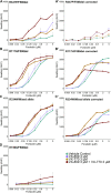
Figure 2.. R347P is functionally unresponsive to VX-770.
Organoids of different patients were treated with an increasing concentration of forskolin, and swelling was measured after 60 min. (A) Nontreated R347P/F508del organoids showed a low amount of swelling, indicative for reduced CFTR function. VX-809 and VX-770 gave a moderate response, and a combination of VX-809 and VX-770 had an additive effect. (A′) Corrected response of A in which 50% of the average swelling value of F508del/F508del organoids from eight patients from Dekkers et al (2016) was subtracted from the values of R347P/F508del. (B) The R117H/F508del showed residual function. VX-770 alone or a combination of VX-770 and VX-809 gave a maximal effect, where a plateau was reached at lower concentrations of forskolin. (B′) R117H/F508del swelling, corrected as in A' (C) R334W/Mixed allele variants with F508del, R746X, and N1303K on the second allele, had a lower but overall similar response as R117H/F508del in B. (C') Same as (A′) and (B′) but subtracted from the R334W/Mixed allele. (D) The F508del/E730X had no residual response and a slight increase upon addition of either drug alone, which was additive to the combination of VX-809 and VX-770. (A–C′) Data represent average ± SD of triplicate measurements of organoids from three distinct patients. Data in (D) represent average ± SD of triplicate measurements of an organoid from one patient.
Figure 3.
Figure 3.. VX-770 does not improve R347P folding.
(A) Cryo-EM structure of human CFTR (Liu et al, 2017) (PBD: 5UAK), generated using Chimera software (Pettersen et al, 2004), illustrating that R117 is in the cytoplasm in the first extracellular loop between TM1 and TM2, R334 is located in the second extracellular loop between TM3 and TM4, and R347 is located in TM6 inside the pore. The colors wheat, purple, gray, green, and blue indicate TMD1, NBD1, R region, TMD2, and NBD2, respectively. (B) CFTR was expressed in HEK293 cells, which were labeled with 35S-methionine/cysteine for 15 min and chased for 0 and 2 h in the presence or absence of 3 μM VX-770. The cells were lysed in 1% Triton X-100 in MNT, and the cell lysates were treated or not with Proteinase K at 25 μg/ml for 15 min. CFTR and fragments were immunoprecipitated using TMD1C (TMD1), Mr. Pink (NBD1 and full-length CFTR), TMD2C (TMD2), and 596 (NBD2) antibodies. * indicates nonspecific bands. Source data are available online for this figure.
Figure 4.
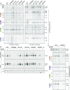
Figure 4.. VX-809 corrects R347P folding.
(A) HEK293 cells expressing CFTR constructs were labeled for 15 min and chased for 0 or 2 h in the presence or absence of VX-809. The cells were lysed in 1% Triton X-100 in MNT, and the cell lysates were incubated with or without Proteinase K at 25 μg/ml for 15 min. CFTR and fragments were immunoprecipitated using domain-specific antibodies as in Fig 3. (B) Cell-surface biotinylation of CFTR in HEK293 cells with or without VX-809 or VX-770 pretreatment. The cells were lysed in 1% Triton X-100 in MNT, and the lysates were used for pull-down of biotinylated proteins with NeutrAvidin beads. 7.5% SDS-PAA gels were run and transferred to PVDF membrane and blotted against CFTR (596) or Actin. (C) Same as in (A) but in the presence of VX-770, VX-809, or both as indicated. All lanes of IP:TMD1 were present on one gel, but the solid black line indicates where the lanes were removed. * indicates nonspecific bands. Source data are available online for this figure.
Figure 5.
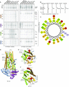
Figure 5.. The proline in R347P destabilizes TM6.
(A) As in Fig 3B, HEK293 cells transiently expressing indicated CFTR constructs were labeled for 15 min and chased for 0 or 2 h. The cells were lysed in 1% Triton X-100 in MNT and incubated with proteinase K for 15 min, followed by immunoprecipitation of CFTR with domain-specific antibodies. The lane labeled C is a nontransfected control. * indicates nonspecific bands. (B) Lane profiles of IP:TMD1 data shown in (A). (C) Helical wheel representation of CFTR residues 331–360, showing that R347 is largely surrounded by small hydrophobic residues. Hydrophilic (circle), hydrophobic (diamond), negatively charged (triangle), positively charged (pentagons). Shades of green indicate the hydrophobicity with dark green having high hydrophobicity and yellow zero hydrophobicity. Hydrophilic residues are shades of red, more intense red means most hydrophilic residue. Potentially charged residues are light blue. (D) Cryo-EM structure of human CFTR illustrating R347 and interacting residues, helices are shown as cylinders (Liu et al, 2017) (PBD: 5UAK). R347 is highlighted in red, and its interacting residues D993 and D924 are colored yellow. Transmembrane helix 6 and the N terminus (AA 1–77) are colored in light blue. Extracellular loops and intracellular loop 1 (ICL1) are labeled in dark gray. The colors wheat, blue, gray, green, and purple indicate TMD1, NBD1, R region, TMD2, and NBD2, respectively. The arrow indicates rotation in relation to Fig 3A left panel. (E) Same as in D, R117 in ECL1 and R334 in ECL2 are shown as red spheres, and their interacting residues E1126 in ECL5 and E217 in ECL3 are represented as yellow spheres. (F) Structural model of CFTR in the open conformation (Mornon et al, 2015), illustrating R117 and R334 in red along with their binding partners in yellow. Colors and annotations are the same as in (D, E). Source data are available online for this figure.

References

    1. Allikmets R, Singh N, Sun H, Shroyer NF, Hutchinson A, Chidambaram A, Gerrard B, Baird L, Stauffer D, Peiffer A, et al. (1997) A photoreceptor cell-specific ATP-binding transporter gene (ABCR) is mutated in recessive Stargardt macular dystrophy. Nat Genet 15: 236–246. 10.1038/ng0397-236 - DOI - PubMed
    1. Braakman I, Hoover-Litty H, Wagner KR, Helenius A (1991) Folding of influenza hemagglutinin in the endoplasmic reticulum. J Cell Biol 114: 401–411. 10.1083/jcb.114.3.401 - DOI - PMC - PubMed
    1. Byrnes LJ, Xu Y, Qiu X, Hall JD, West GM (2018) Sites associated with Kalydeco binding on human cystic fibrosis transmembrane conductance regulator revealed by hydrogen/deuterium exchange. Sci Rep 8: 4664 10.1038/s41598-018-22959-6 - DOI - PMC - PubMed
    1. Castellani C, Cuppens H, Macek M Jr., Cassiman JJ, Kerem E, Durie P, Tullis E, Assael BM, Bombieri C, Brown A, et al. (2008) Consensus on the use and interpretation of cystic fibrosis mutation analysis in clinical practice. J Cyst Fibros 7: 179–196. 10.1016/j.jcf.2008.03.009 - DOI - PMC - PubMed
    1. Chen PC, Olson EM, Zhou Q, Kryukova Y, Sampson HM, Thomas DY, Shyng SL (2013) Carbamazepine as a novel small molecule corrector of trafficking-impaired ATP-sensitive potassium channels identified in congenital hyperinsulinism. J Biol Chem 288: 20942–20954. 10.1074/jbc.m113.470948 - DOI - PMC - PubMed

Publication types

MeSH terms

Associated data