. 2019 Mar 22;294(12):4621-4633.
doi: 10.1074/jbc.RA118.006032. Epub 2019 Jan 18.

A single discrete Rab5-binding site in phosphoinositide 3-kinase β is required for tumor cell invasion

Affiliations

A single discrete Rab5-binding site in phosphoinositide 3-kinase β is required for tumor cell invasion

Samantha D Heitz et al. J Biol Chem. .

Abstract

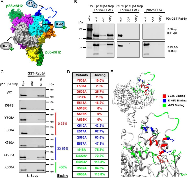
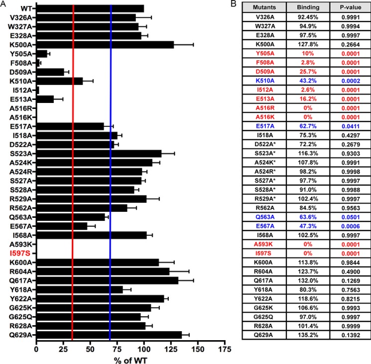
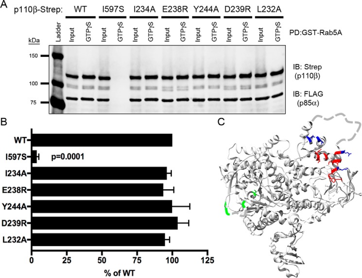
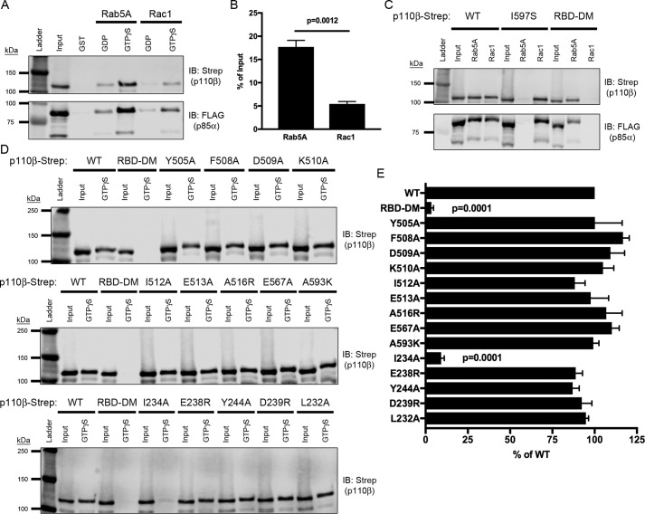
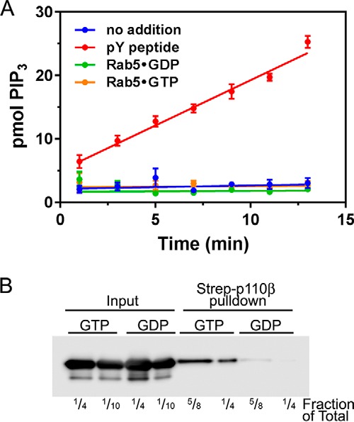
Phosphoinositide 3-kinase β (PI3Kβ) is regulated by receptor tyrosine kinases (RTKs), G protein-coupled receptors (GPCRs), and small GTPases such as Rac1 and Rab5. Our lab previously identified two residues (Gln596 and Ile597) in the helical domain of the catalytic subunit (p110β) of PI3Kβ whose mutation disrupts binding to Rab5. To better define the Rab5-p110β interface, we performed alanine-scanning mutagenesis and analyzed Rab5 binding with an in vitro pulldown assay with GST-Rab5GTP Of the 35 p110β helical domain mutants assayed, 11 disrupted binding to Rab5 without affecting Rac1 binding, basal lipid kinase activity, or Gβγ-stimulated kinase activity. These mutants defined the Rab5-binding interface within p110β as consisting of two perpendicular α-helices in the helical domain that are adjacent to the initially identified Gln596 and Ile597 residues. Analysis of the Rab5-PI3Kβ interaction by hydrogen-deuterium exchange MS identified p110β peptides that overlap with these helices; no interactions were detected between Rab5 and other regions of p110β or p85α. Similarly, the binding of Rab5 to isolated p85α could not be detected, and mutations in the Ras-binding domain (RBD) of p110β had no effect on Rab5 binding. Whereas soluble Rab5 did not affect PI3Kβ activity in vitro, the interaction of these two proteins was critical for chemotaxis, invasion, and gelatin degradation by breast cancer cells. Our results define a single, discrete Rab5-binding site in the p110β helical domain, which may be useful for generating inhibitors to better define the physiological role of Rab5-PI3Kβ coupling in vivo.

Keywords: G protein-coupled receptor (GPCR); GTPase; PIK3CB; PIK3R1; Rab; breast cancer; chemotaxis; invasion; matrix degradation; p85; phosphoinositide 3-kinase (PI 3-kinase); receptor tyrosine kinase.


Conflict of interest statement

J. M. B. is on the scientific advisory board of Karus Therapeutics

Figures

Figure 1.
Figure 1.
Mutagenesis of the p110β helical domain disrupts Rab5A binding to PI3Kβ. A, space-filling model of the murine p110β catalytic subunit with the iSH2 and cSH2 domains of the p85β regulatory subunit (green) (Protein Data Bank (PDB) code 2Y3A). Yellow, the N-terminal ABD; gray, the RBD; orange, the C2 domain; cyan, the helical domain; purple, the C-terminal kinase domain. The blue dashed line represents the C2–helical linker, which was not observed in the X-ray structure. The arrows indicate where Rac1, Rab5, and Gβγ bind to p110β. Gln596/Ile597, whose mutation disrupts Rab5 binding, are shown in white. B, representative immunoblots (IB) of the GST-Rab5A pulldown (PD) assay. Human GST-Rab5A was immobilized on GSH-agarose beads and loaded with GDP or GTPγS. The beads were incubated with whole-cell lysates (Input) from HEK293T cells transfected with p85α-FLAG without or with WT p110β-Strep or the Rab5-uncoupled I597S mutant. C, representative immunoblots of the GST-Rab5A pulldown assay with p110β mutants. Samples were analyzed by SDS-PAGE and blotted for Strep (p110β) and FLAG (p85α). D, table of Rab5A-binding activity for representative p110β mutants. Binding to GTPγS–Rab5A was calculated as a percentage of the input and then normalized to WT p110β binding, which was set to 100%. Binding, as compared with WT p110β, was stratified into three groups: 0–33% (red), 33–66% (blue), and >66% (green). Residues in the Gβγ-binding loop are indicated with an asterisk. E, ribbon diagrams of p110β (upper panel) and a magnified view of the helical domain (lower panel). Residues are color-coded based on their Rab5A-binding activity (PBD code 2Y3A).
Figure 2.
Figure 2.
Helical domain mutations in p110β exhibit variable binding to GST-Rab5A. A, quantification of the Rab5A-binding activity for all p110β mutants as compared with WT. Data represent the mean ± S.E. from three independent experiments. Error bars represent S.E. The red and blue lines indicate 33 and 66% binding, respectively. B, table showing the average Rab5A-binding activity for all p110β mutants relative to WT p110β. Red, 0–33%; blue, 33–66%. Statistical analyses were performed using one-way ANOVA. Residues in the Gβγ-binding loop are indicated with an asterisk.
Figure 3.
Figure 3.
Mutation of the p110β RBD does not affect binding to Rab5. A, representative immunoblot (IB) of GST-Rab5A pulldown (PD) assay showing lysates from cells expressing select RBD p110β mutations incubated with GST-Rab5A beads and blotted for Strep and FLAG. B, quantification of Rab5A binding. The data represent the mean ± S.E. from three independent experiments. Error bars represent S.E. Statistical analyses were performed using one-way ANOVA. No statistical differences were observed between WT and RBD p110β mutant proteins. C, ribbon diagram of p110β showing residues in the helical domain and RBD that were targeted for mutagenesis. Color coding reflects Rab5-binding activity relative to WT p110β: red, 0–33%; blue: 33–66%; green, >66%. Residues in the RBD and Gβγ-binding loop that are not observed in the X-ray structure are depicted as dashed lines.
Figure 4.
Figure 4.
Mutation of the Rab5-binding interface does not disrupt binding to Rac1. A, representative immunoblot (IB) of GST-Rab5A and GST-Rac1 pulldown assay. Lysates expressing WT p110β-Strep/p85α-FLAG were incubated with GST-Rab5A and GST-Rac1 beads loaded with nucleotide and assessed for binding via SDS-PAGE and immunoblotting for Strep and FLAG. B, quantification of PI3Kβ binding to GTPγS–Rac1 or Rab5A, expressed as a percentage of the input. Data represent the mean ± S.E. from three independent experiments. Statistical analysis was performed using an unpaired Student's t test. C, representative immunoblot of GTPγS-loaded GST-Rab5A and GST-Rac1 pulldowns incubated with lysates expressing p85α-FLAG and WT, I597S, or Rac1-uncoupled mutant (RBD-DM) p110β-Strep. D, GST-Rac1 binding assay with lysates expressing Rab5-uncoupled helical domain and RBD p110β mutants analyzed by SDS-PAGE and immunoblotted with Strep. E, quantification of the percent binding as compared with WT p110β. Data represent the mean ± S.E. from three independent experiments. Error bars represent S.E. in all panels. Statistical analyses were performed using one-way ANOVA. No statistically significant difference was observed, unless indicated.
Figure 5.
Figure 5.
Mutation of the Rab5-binding interface does not affect PI3Kβ kinase activity or activation by Gβγ. A, ribbon diagram of the p110β helical domain showing the original Rab5-uncoupled mutations (Gln596/Ile597) and mutated residues selected for in vitro kinase assays. Color coding corresponds to relative Rab5-binding activity: residues Phe508 and Ile512 (red), 0–33%; Lys510 and Glu517 (blue), 33–66%. B, quantification of in vitro kinase activity for WT p110β, KD (K805R) p110β, and the four helical domain p110β mutants. Values were normalized to the amount of p110β in each reaction as determined by immunoblotting. Data represent the mean ± S.E. for four independent experiments. Statistical analyses were performed using one-way ANOVA. Significance was observed for the difference in kinase activity between WT and KD (p = 0.0097), but no significant difference was observed between WT and other mutants of p110β. C, quantification of in vitro kinase activity without (black bars) and with (white bars) Gβγ, normalized to unstimulated WT p110β activity. Data represent the mean ± S.E. for three independent experiments. Statistical analyses were performed using an unpaired Student's t test. D, quantification of the -fold change in kinase activity with Gβγ stimulation. Data represent the mean ± S.E. for three independent experiments. Statistical analyses were performed using one-way ANOVA. There was no significant difference in the -fold activation for WT and mutant p110β. E, activation of WT and Gβγ-uncoupled (K532D/K533D (KKDD)) PI3Kβ by Gβγ was determined as in D. The data are the mean ± S.D. from two experiments. Error bars represent S.E. in panels B, C and D, and S.D. in panel E.
Figure 6.
Figure 6.
HDX-MS revealed changes within the helical domain of p110β upon interaction with soluble Rab5A. HDX-MS experiments were carried out for PI3Kβ in the absence or presence of a 7.5-fold molar excess of soluble Rab5A. A, left panel, representation of the p110β helical domain with Rab5-uncoupled mutants color-coded by relative Rab5 binding as compared with WT. Right panel, representation of the p110β helical domain showing peptides that exhibited changes in deuteration in the presence of Rab5A. B, representation of the full-length p110β showing all peptides that exhibited significant changes in deuteration in the presence of Rab5A (>5% and 0.5-Da difference in deuterium incorporation and p value of <0.01 based on a Student's t test). C, time course of deuterium incorporation for select peptides in the absence (solid black line) or presence (dashed line) of Rab5A. Data represent the mean ± S.E. for three independent experiments. For most data points the error bars, which represent S.E., are contained within the symbols.
Figure 7.
Figure 7.
Rab5GTP does not affect PI3Kβ kinase activity in vitro. Purified recombinant PI3Kβ was incubated with 10 μm Rab5, which had been loaded with GDP or GTPγS, or 1 μm tyrosine bisphosphopeptide (pY peptide). A, lipid kinase activity toward lipid vesicles (2.9 mol % PIP2) was determined as described. The data are the mean ± S.E. from three independent experiments. Error bars represent S.E. B, p85-FLAG/p110β-StrepII was produced in HEK293T cells and immobilized on Strep-Tactin beads. The beads were incubated with GDP- or GTP-loaded Rab5, washed, and then analyzed by SDS-PAGE and Western blotting with Rab5 antibodies. The lanes show ¼ or 110 of the input and ⅝ or ¼ of the pulldowns.
Figure 8.
Figure 8.
Rab5 binding to p110β is required for chemotaxis, invasion, and gelatin degradation by breast cancer cells. A, myc immunoprecipitation and p85 blotting of p110β knockdown MDA-MB-231 cells stably expressing murine WT, Gβγ-uncoupled (K526D/K527D (KKDD)), kinase-dead (K799R (KR)), or Rab5-uncoupled p110β (Q590C (QC) or I591S (IS)). B, LPA-stimulated (10 μm) chemotaxis of MDA-MB-231 p110β knockdown cells stably expressing murine WT p110β or the Rab5-uncoupled p110β mutant (IS). Data represent the mean ± S.E. from three independent experiments. C, EGF-stimulated (5 nm) chemotaxis of MDA-MB-231 p110β knockdown cells stably expressing murine WT p110β or the Rab5-uncoupled p110β mutant (I591S). Data represent the mean ± S.E. from three independent experiments. D, transwell Matrigel invasion assay toward 5 nm EGF of MDA-MB-231 p110β knockdown cells stably expressing WT or I591S p110β. The data were normalized to the number of invaded cells expressing WT p110β and represent the mean ± S.E. for three independent experiments. E, MDA-MB-231 cells expressing WT or I591S p110β were plated on Oregon Green 488–conjugated gelatin for 18 h, fixed, and stained with rhodamine phalloidin. Scale bar = 20 μm. F, degradation area per cell for MDA-MB-231 p110β knockdown cells stably expressing WT or I591S p110β. Error bars represent S.E. in all panels. Statistical analyses were performed using one-way ANOVA (B, C, and D) or Student's t test (E).

References

    1. Vanhaesebroeck B., Leevers S. J., Ahmadi K., Timms J., Katso R., Driscoll P. C., Woscholski R., Parker P. J., and Waterfield M. D. (2001) Synthesis and function of 3-phosphorylated inositol lipids. Annu. Rev. Biochem. 70, 535–602 10.1146/annurev.biochem.70.1.535 - DOI - PubMed
    1. Vanhaesebroeck B., Guillermet-Guibert J., Graupera M., and Bilanges B. (2010) The emerging mechanisms of isoform-specific PI3K signalling. Nat. Rev. Mol. Cell Biol. 11, 329–341 10.1038/nrm2882 - DOI - PubMed
    1. Zhang X., Vadas O., Perisic O., Anderson K. E., Clark J., Hawkins P. T., Stephens L. R., and Williams R. L. (2011) Structure of lipid kinase p110β/p85β elucidates an unusual SH2-domain-mediated inhibitory mechanism. Mol. Cell 41, 567–578 10.1016/j.molcel.2011.01.026 - DOI - PMC - PubMed
    1. Kurosu H., Maehama T., Okada T., Yamamoto T., Hoshino S., Fukui Y., Ui M., Hazeki O., and Katada T. (1997) Heterodimeric phosphoinositide 3-kinase consisting of p85 and p110β is synergistically activated by the βγ subunits of G proteins and phosphotyrosyl peptide. J. Biol. Chem. 272, 24252–24256 10.1074/jbc.272.39.24252 - DOI - PubMed
    1. Dbouk H. A., Vadas O., Shymanets A., Burke J. E., Salamon R. S., Khalil B. D., Barrett M. O., Waldo G. L., Surve C., Hsueh C., Perisic O., Harteneck C., Shepherd P. R., Harden T. K., Smrcka A. V., et al. (2012) G protein-coupled receptor-mediated activation of p110β by Gβγ is required for cellular transformation and invasiveness. Sci. Signal. 5, ra89 10.1126/scisignal.2003264 - DOI - PMC - PubMed

Publication types

Associated data