. 2019 Mar 29;294(13):4815-4827.
doi: 10.1074/jbc.RA118.005739. Epub 2019 Jan 18.

Hepatitis B virus X protein-induced SH2 domain-containing 5 (SH2D5) expression promotes hepatoma cell growth via an SH2D5-transketolase interaction

Affiliations

Hepatitis B virus X protein-induced SH2 domain-containing 5 (SH2D5) expression promotes hepatoma cell growth via an SH2D5-transketolase interaction

Yongfa Zheng et al. J Biol Chem. .

Abstract

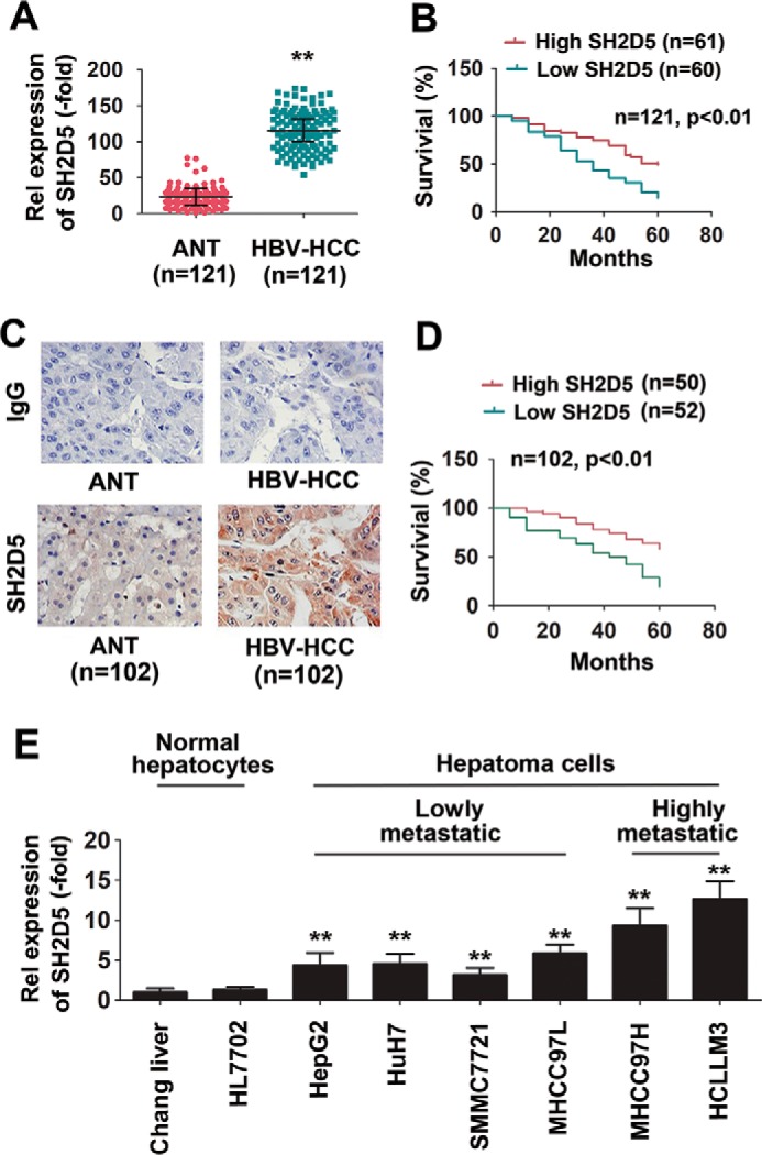
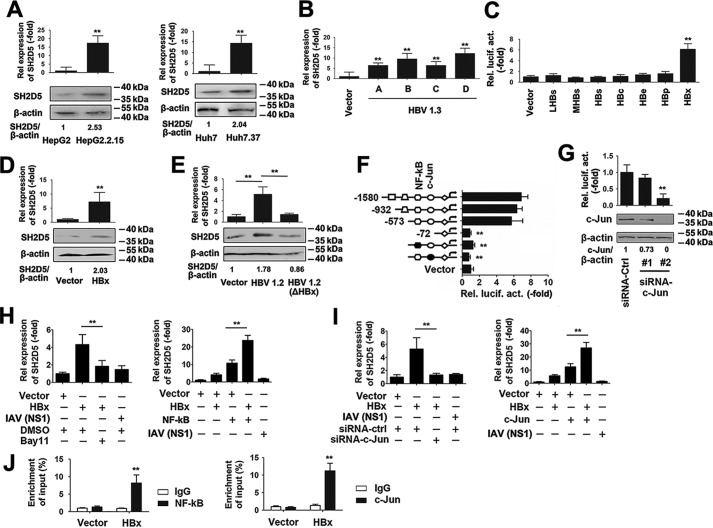
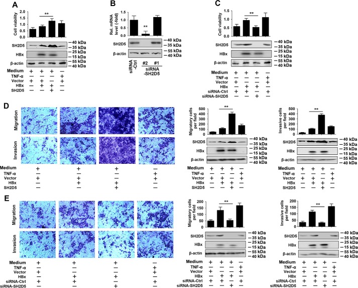
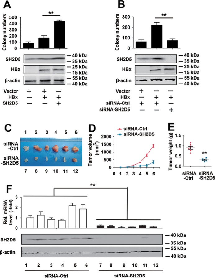
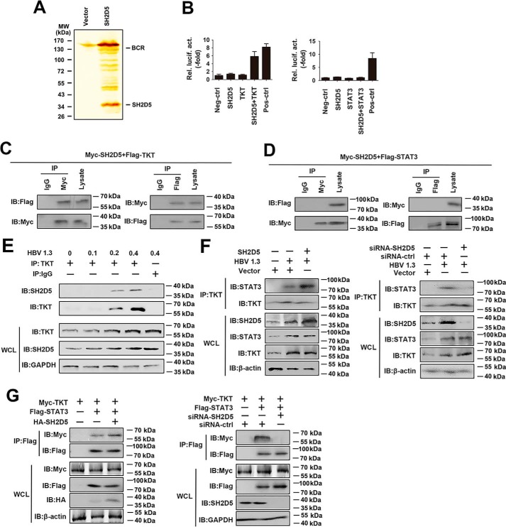
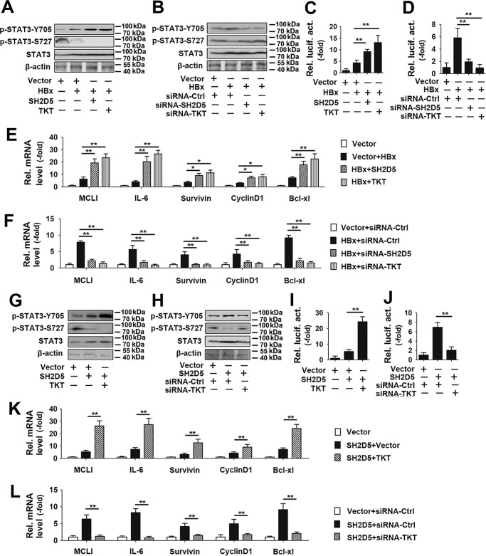
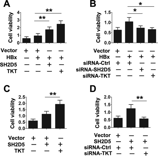
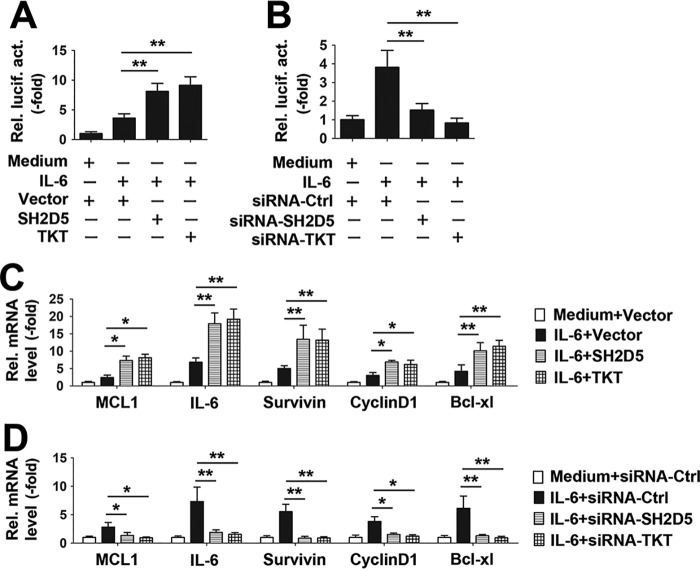
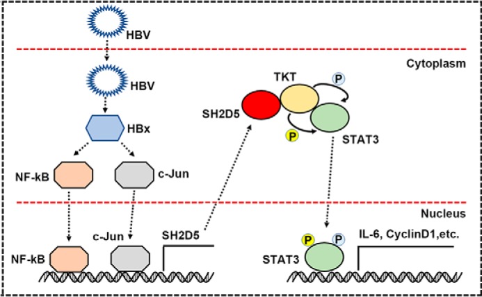
Hepatitis B virus X protein (HBx) critically contributes to the development of hepatocellular carcinoma (HCC). However, the mechanisms by which HBx promotes HCC remain unclear. In the present study, using a combination of gene expression profiling and immunohistochemistry, we found higher levels of SH2 domain-containing 5 (SH2D5) in liver tissue from HBV-associated HCC (HBV-HCC) patients than in adjacent nontumor tissues. Moreover, HBV infection elevated SH2D5 levels, and we observed that HBx plays an important role in SH2D5 induction. We also found that HBx triggers SH2D5 expression through the NF-κB and c-Jun kinase pathways. Employing SH2D5 overexpression or knockdown, we further demonstrate that SH2D5 promotes HCC cell proliferation both in vitro and in vivo While investigating the mechanism of SH2D5-mediated stimulation of HCC cell proliferation, we noted that HBV induces SH2D5 binding to transketolase (TKT), a pentose phosphate pathway enzyme, thereby promoting an interaction between and signal transducer and activator of transcription 3 (STAT3). Furthermore, HBx stimulated STAT3 phosphorylation at Tyr-705 and promoted the activity and downstream signaling pathway of STAT3 via the SH2D5-TKT interaction. Taken together, our results suggest that SH2D5 is an HBV-induced protein capable of binding to TKT, leading to induction of HCC cell proliferation.

Keywords: HBV X protein; HBV-associated HCC; IL-6-STAT3 signaling; SH2D5; cancer biology; cell migration; hepatitis B virus (HBV, Hep B); inflammation; interleukin 6 (IL-6); transketolase.


Conflict of interest statement

The authors declare that they have no conflicts of interest with the contents of this article

Figures

Figure 1.
Figure 1.
SH2D5 is increased in HCC and is clinically relevant to patient prognosis. A, real-time PCR assays of SH2D5 expression levels in HCC tissues and their corresponding ANT. The lowest value of ANT was designated as 1. SH2D5 data are expressed as fold induction (-fold) relative to the lowest value of healthy individuals. Data represent means ± S.E. B, Kaplan-Meier plots of HCC patients stratified by SH2D5 RNA levels. The median level of SH2D5 expression in each panel was used as the cutoff, with log-rank test for significance. C, immunohistochemical staining of SH2D5 in HCC tissues and their corresponding ANT. IgG was use as isotype control antibody. D, Kaplan-Meier plots of HCC patients stratified by SH2D5 protein levels. The median level of SH2D5 expression in each panel was used as the cutoff, with log-rank test for significance. E, real-time PCR analysis of SH2D5 expression in human normal hepatocytes and HCC cell lines. All experiments were repeated at least three times. (**, p < 0.01).
Figure 2.
Figure 2.
SH2D5 was induced by HBx at the transcriptional level. A, SH2D5 mRNA levels and protein levels in HepG2 and HepG2.2.15 cells (left panel) and Huh7 and Huh7.37 cells (right panel). B, HepG2 cells were transfected with vector control or pHBV-1.3 (genotype A-,-D) for 48 h prior to real-time PCR assays. C, HepG2 cells were co-transfected with pSH2D5-Luc and the indicated plasmids with viral protein-coding sequences for 48 h prior to luciferase activity reporter assays. D, HepG2 cells were transfected with the vector control or pCMV-HBx for 48 h prior to real-time PCR (upper panel) and Western blotting (lower panel) analyses. E, Huh7 cells were transfected with vector control, pHBV-1.2, or pHBV-1.2 (ΔHBx), an HBx-deficient HBV mutant for 48 h. SH2D5 expression was quantified prior to real-time PCR (upper panel) and Western blotting (lower panel) analyses. F, schematic diagram of individual SH2D5 promoter cis-regulatory elements and SH2D5-truncated or site-specific mutants (left), and the results are from luciferase activity assays (right). Huh7 cells were transfected with the indicated plasmids for 48 h prior to luciferase assays. G, experiments were performed similar to those in D, except c-Jun–specific siRNA (siRNA–c-Jun) was used. H, Huh7 cells were transfected with indicated plasmids and treated with or without NF-κB inhibitor Bay11-7082 (left panel) or transfected with vector control, pCMV-HBx, pCMV-NF-κB, or pCMV-NS1 of IAV (right panel) for 48 h, respectively. SH2D5 RNA levels were quantified by real-time PCR assays. I, experiments were performed similar to those in H, except siRNA–c-Jun (left panel) or pCMV–c-Jun (right panel) was used. J, Huh7 cells were transfected with vector control or pCMV-HBx for 48 h. ChIP assays were performed with anti-NF-κB (left panel), anti-c-Jun (right panel), or IgG-conjugated agarose. Promoter sequences in the input DNA and the DNA recovered from antibody-bound chromatin segments were detected using real-time PCR. Enrichment was determined relative to input controls. In the real-time RT-PCR experiments, the control was designated 1. In the Western blotting experiments, intensities of bands were analyzed by ImageJ and normalized to the corresponding controls. All experiments were repeated at least three times. Bar graphs present means ± S.D., n = 3 (**, p < 0.01).
Figure 3.
Figure 3.
SH2D5 promoted HCC cell proliferation, migration, and invasion in vitro. A, HepG2 cells were transfected with the indicated plasmids for 24 h and treated with or without TNFα (50 ng/ml) for 48 h prior to cell proliferation (upper panel) and Western blotting assays (lower panel). B, Huh7 cells were transfected with siRNA control or siRNA-SH2D5s for 48 h prior to real-time PCR (upper panel) and Western blotting (lower panel) analyses. C, experiments were performed similar to those in A, except the indicated siRNA-SH2D5 #2 was used. D, HepG2 cells were transfected with the indicated plasmids and treated with or without TNFα (50 ng/ml) for 24 h, after culture in a Transwell (upper panel) or invasion (lower panel) system for 24 h. E, experiments were performed similar to those in D, except the indicated siRNA-SH2D5 #2 was used. In the real-time RT-PCR experiments, the control was designated 1. All experiments were repeated at least three times. Bar graphs present means ± S.D., n = 3 (**, p < 0.01).
Figure 4.
Figure 4.
SH2D5 promoted proliferation of hepatoma cells in vivo. A, HepG2 cells were transfected with vector control, pCMV-HBx, or pCMV-SH2D5 and seeded into the soft agar dish. Colonies under the microscope were counted after a 4-week incubation (upper panel). SH2D5 and HBx expressions were quantified by Western blot assays (lower panel). B, experiments were performed similar to those in A, except the indicated siRNA-SH2D5 was used. C, nude mice were sacrificed and photographed after 1 month of subcutaneous injection. D, growth curves of tumors derived from HepG2-X cells transfected with si-SH2D5 or si-Ctrl. E, average weight of tumors. F, relative mRNA and protein levels of SH2D5 in the tumor tissues from mice were detected by real-time PCR (upper panel) and Western blot assays (lower panel). All experiments were repeated at least three times. Bar graphs present means ± S.D., n = 3 (**, p < 0.01).
Figure 5.
Figure 5.
HBV promotes the interaction of SH2D5 and TKT, and the subsequent recruitment of STAT3. A, HepG2 cells were transfected with vector control or FLAG-SH2D5 for 48 h. Cells were lysed and were immunopurified with anti-FLAG affinity columns and eluted with FLAG peptide. The eluates were resolved by SDS-PAGE and silver-stained. The differential protein bands were retrieved and analyzed by MS. B, determination of SH2D5 and TKT interaction in 293T cells by mammalian two-hybrid analysis. 293T cells were co-transfected with control plasmids, pG5-luc (a luciferase reporter plasmid), pVP16-SH2D5, pM-TKT (left panel), or pM-STAT3 (right panel). Interaction between proteins was monitored by luciferase activity, which was measured 48 h after transfection. C, 293T cells were transfected with FLAG-tagged TKT (FLAG-TKT) and Myc-tagged SH2D5 (Myc-SH2D5). Forty eight hours post-transfection, co-immunoprecipitation (IP) and immunoblot (IB) analyses were performed with the indicated antibodies. D, experiments were performed similar to those in C, except FLAG-STAT3 was used. E, HepG2 cells were transfected with increasing amounts of HBV 1.3 plasmid. Forty eight hours post-transfection, co-immunoprecipitation and immunoblot analyses were performed with the indicated antibodies. F, experiments were performed similar to those in E, except SH2D5 plasmid (left panel) or si-SH2D5 (right panel) was used. G, 293T cells were transfected with the indicated plasmids or siRNAs. Forty eight hours post-transfection, co-immunoprecipitation and immunoblot analyses were performed with the indicated antibodies. All experiments were repeated at least three times. WCL, whole-cell lysates.
Figure 6.
Figure 6.
Effect of SH2D5 and TKT on HBx regulated phosphorylation and downstream signaling of STAT3. A, HepG2 cells were transfected with the indicated plasmids for 24 h prior to Western blotting analyses. B, experiments were performed similar to those in A, except the indicated siRNA-TKT was used. C, HepG2 cells were transfected with the indicated plasmids for 36 h prior to luciferase activity assays. D, experiments were performed similar to those in C, except the indicated siRNA-TKT was used. E, HepG2 cells transfected with the indicated plasmids for 36 h prior to real-time PCR analyses. F, experiments were performed similar to those in E, except the indicated siRNA-TKT was used. G and H, experiments were performed similar to those in A and B, except without HBx plasmid. I and J, experiments were performed similar to those in C and D, except without HBx plasmid. K and L, experiments were performed similar to those in E and F, except without HBx plasmid. All experiments were repeated at least three times. Bar graphs present means ± S.D., n = 3 (**, p < 0.01; *, p < 0.05).
Figure 7.
Figure 7.
Verification of the interaction of SH2D5 and TKT in HCC cells proliferation. A, HepG2 cells were transfected with vector control, pCMV-HBx, pCMV-TKT, or pCMV-SH2D5 for 3 days prior to cell proliferation assays. B, HepG2 cells were transfected with vector control, pCMV-HBx, or indicated siRNAs for 3 days prior to cell proliferation assays. C, HepG2 cells were transfected with vector control, pCMV-TKT, or pCMV-SH2D5 for 3 days prior to cell proliferation assays. D, HepG2 cells were transfected with vector control, pCMV-SH2D5, siRNA control, or siRNA-TKT for 3 days prior to cell proliferation assays. All experiments were repeated at least three times for consistent results. Bar graphs present means ± S.D., n = 3 (**, p < 0.01; *, p < 0.05).
Figure 8.
Figure 8.
Verification of the role of SH2D5 and TKT in IL-6–STAT3 signaling pathway. A, HepG2 cells transfected with the indicated plasmids for 12 h and treated with or without IL-6 (10 ng/ml) for 12 h prior to luciferase assays. B, HepG2 cells transfected with the indicated siRNAs for 12 h and treated with or without IL-6 (10 ng/ml) for 12 h prior to luciferase assays. C and D, experiments were performed similar to those in A and B, except the downstream genes in the JAK–STAT3 pathway were detected by real-time PCR. All experiments were repeated at least three times for consistent results. Bar graphs present means ± S.D., n = 3 (**, p < 0.01; *, p < 0.05).
Figure 9.
Figure 9.
Hypothetical model for SH2D5 induction following HBV infection and the role of SH2D5 in the progression of HBV-HCC.

Similar articles

Cited by

References

    1. Chen S., Dong Z., Yang P., Wang X., Jin G., Yu H., Chen L., Li L., Tang L., Bai S., Yan H., Shen F., Cong W., Wen W., and Wang H. (2017) Hepatitis B virus X protein stimulates high mobility group box 1 secretion and enhances hepatocellular carcinoma metastasis. Cancer Lett. 394, 22–32 10.1016/j.canlet.2017.02.011 - DOI - PubMed
    1. Hsieh A., Kim H. S., Lim S. O., Yu D. Y., and Jung G. (2011) Hepatitis B viral X protein interacts with tumor suppressor adenomatous polyposis coli to activate Wnt/β-catenin signaling. Cancer Lett. 300, 162–172 10.1016/j.canlet.2010.09.018 - DOI - PubMed
    1. Torre L. A., Bray F., Siegel R. L., Ferlay J., Lortet-Tieulent J., and Jemal A. (2015) Global cancer statistics, 2012. CA Cancer J. Clin. 65, 87–108 10.3322/caac.21262 - DOI - PubMed
    1. Yang P., Li Q. J., Feng Y., Zhang Y., Markowitz G. J., Ning S., Deng Y., Zhao J., Jiang S., Yuan Y., Wang H. Y., Cheng S. Q., Xie D., and Wang X. F. (2012) TGF-β-miR-34a-CCL22 signaling-induced Treg cell recruitment promotes venous metastases of HBV-positive hepatocellular carcinoma. Cancer Cell 22, 291–303 10.1016/j.ccr.2012.07.023 - DOI - PMC - PubMed
    1. Liu D., Cui L., Wang Y., Yang G., He J., Hao R., Fan C., Qu M., Liu Z., Wang M., Chen L., Li H., and Guo D. (2016) Hepatitis B e antigen and its precursors promote the progress of hepatocellular carcinoma by interacting with NUMB and decreasing p53 activity. Hepatology 64, 390–404 10.1002/hep.28594 - DOI - PubMed

Publication types

MeSH terms