. 2019 Feb 25;132(5):jcs222018.
doi: 10.1242/jcs.222018.

Recruitment of Jub by α-catenin promotes Yki activity and Drosophila wing growth

Affiliations

Recruitment of Jub by α-catenin promotes Yki activity and Drosophila wing growth

Herve Alégot et al. J Cell Sci. .

Abstract

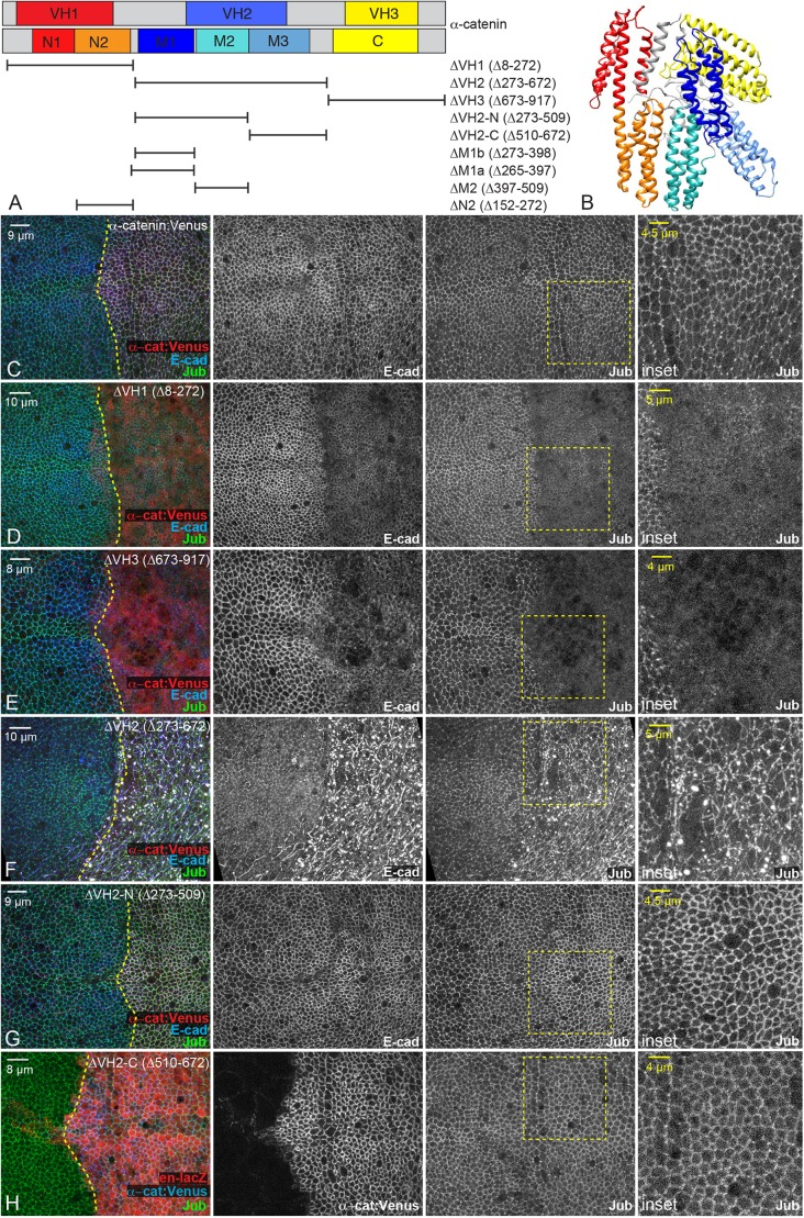
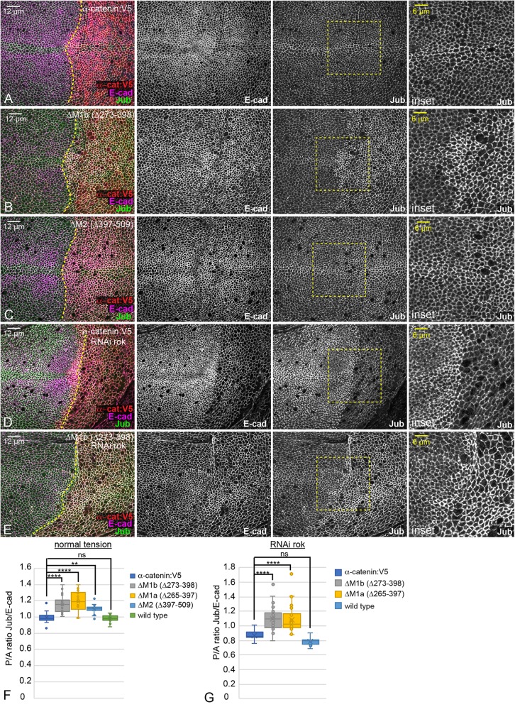
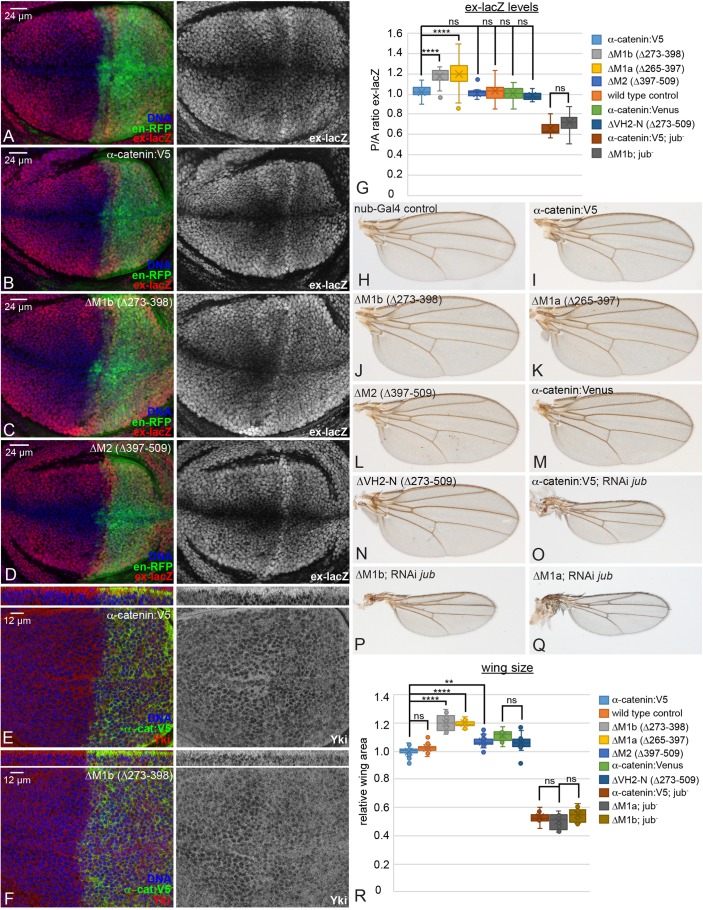
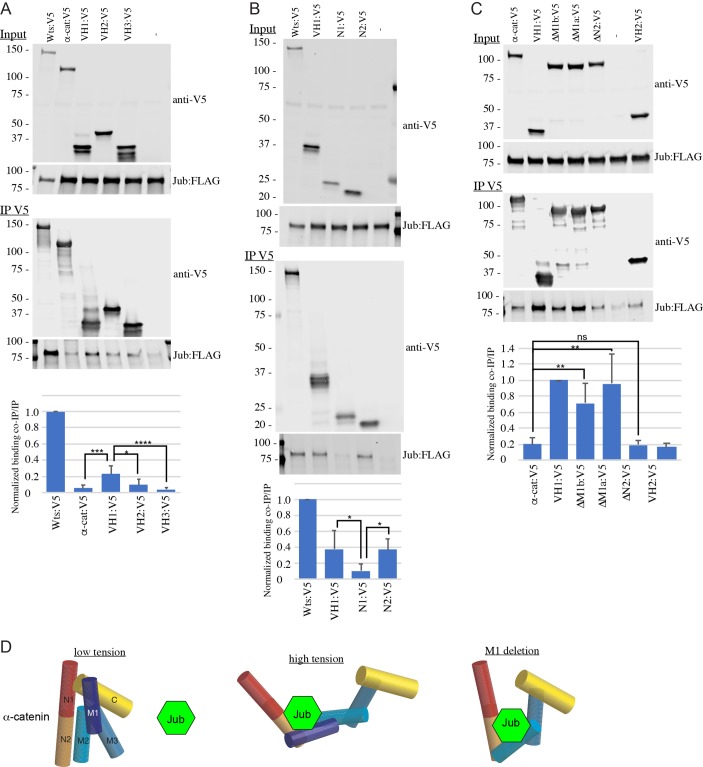
The Hippo signaling network controls organ growth through YAP family transcription factors, including the Drosophila Yorkie protein. YAP activity is responsive to both biochemical and biomechanical cues, with one key input being tension within the F-actin cytoskeleton. Several potential mechanisms for the biomechanical regulation of YAP proteins have been described, including tension-dependent recruitment of Ajuba family proteins, which inhibit kinases that inactivate YAP proteins, to adherens junctions. Here, we investigate the mechanism by which the Drosophila Ajuba family protein Jub is recruited to adherens junctions, and the contribution of this recruitment to the regulation of Yorkie. We identify α-catenin as the mechanotransducer responsible for tension-dependent recruitment of Jub by identifying a region of α-catenin that associates with Jub, and by identifying a region, which when deleted, allows constitutive, tension-independent recruitment of Jub. We also show that increased Jub recruitment to α-catenin is associated with increased Yorkie activity and wing growth, even in the absence of increased cytoskeletal tension. Our observations establish α-catenin as a multi-functional mechanotransducer and confirm Jub recruitment to α-catenin as a key contributor to biomechanical regulation of Hippo signaling.

Keywords: Ajuba; Growth; Hippo; Mechanotransduction; Tension.


Conflict of interest statement

Competing interestsThe authors declare no competing or financial interests.

Figures

Fig. 1.
Fig. 1.
Influence of α-catenin deletions on Jub. (A) Schematics of α-catenin and constructs, with VH regions colored at top and helical bundles colored below. Black bars indicate deletions in constructs, named at right. Amino acids deleted in each construct are indicated within parentheses. (B) Predicted structure of Drosophila α-catenin. Helical bundles are colored as in A. (C-H) Images of wing discs showing localization of E-cadherin (E-cad), Jub and a marker for posterior cells as indicated. UAS-RNAi α-catenin and a Venus-tagged UAS-α-catenin rescue construct were co-expressed under en-Gal4 control. Yellow dashed line indicates the boundary of en-Gal4 expression; boxed area in each third image indicates higher magnification images shown at the far right (inset). (C) Full-length α-catenin. (D) ΔVH1 and tub-Gal80ts, shifted to 29°C for 30 h. (E) ΔVH3 and tub-Gal80ts, shifted to 29°C for 30 h. (F) ΔVH2. (G) ΔVH2-N. (H) ΔVH2-C.
Fig. 2.
Fig. 2.
M1 deletions allow Jub recruitment to AJs under low tension. (A-E) Images of wing discs showing localization of E-cadherin, Jub, and V5-tagged α-catenin. UAS-RNAi α-catenin and UAS-α-catenin constructs were co-expressed under en-Gal4 control. (A) Full-length α-catenin. (B) ΔM1b. (C) ΔM2. (D) Full-length α-catenin and UAS-RNAi-rok. (E) ΔM1b and UAS-RNAi-rok. Yellow dashed line indicates the boundary of en-Gal4 expression; boxed area in each third image indicates higher magnification images shown at the far right (inset). (F) Quantification of Jub (normalized to E-cadherin) in posterior cells as compared to anterior cells, in discs expressing the indicated constructs, displayed as a Tukey box plot, with × marking the mean. n=15 (full length), 20 (ΔM1b), 14 (ΔM1a), 18 (ΔM2), 11 (wt). (G) Quantification of Jub (normalized to E-cadherin) in posterior cells expressing UAS-RNAi-rok as compared to anterior cells, in discs expressing the indicated constructs, displayed as a Tukey box plot, with × marking the mean. n=24 (full length), 31 (ΔM1b), 30 (ΔM1a), 12 (wt).
Fig. 3.
Fig. 3.
Influence of α-catenin deletions on Yki activity. (A-D) Images of wing discs stained for DNA, ex-lacZ, and a marker for posterior cells. UAS-RNAi α-catenin and UAS-α-catenin rescue constructs were co-expressed under en-Gal4 control. (A) UAS-RFP control. (B) Full-length α-catenin. (C) ΔM1b. (D) ΔM2. (E,F) Images of wing discs stained for DNA, Yki and a marker for posterior cells. UAS-RNAi α-catenin and a UAS-α-catenin rescue construct were co-expressed under en-Gal4 control. Thin panels above each image show vertical sections. (E) Full-length α-catenin. (F) ΔM1b. (G) Quantification of ex-lacZ in posterior cells as compared to anterior cells, displayed as a Tukey box plot, with × marking the mean. n=19 (V5-tagged full length), 18 (ΔM1b), 31 (ΔM1a), 12 (ΔM2), 26 (wt), 30 (Venus-tagged full length), 13 (ΔVH2-N), 10 (jub RNAi), 10 (ΔM1b with jub RNAi). (H-Q) Adult wings from flies expressing UAS-α-catenin constructs under nub-Gal4 control. (H) control. (I) Full-length V5-tagged α-catenin. (J) ΔM1b. (K) ΔM1a. (L) ΔM2. (M) Full-length Venus-tagged α-catenin. (N) ΔVH2-N. (O) Full-length V5-tagged α-catenin plus UAS-RNAi-jub. (P) ΔM1b plus UAS-RNAi-jub. (Q) ΔM1a plus UAS-RNAi-jub. (R) Quantification of relative wing areas, displayed as a Tukey box plot, with × marking the mean. n=21 (control), 30 (V5-tagged full length), 12 (ΔM1b), 19 (ΔM1a, ΔM2), 14 (Venus-tagged full length), 20 (ΔVH2-N), 11(jub RNAi), 11 (ΔM1a with jub RNAi), 14 (ΔM1b with jub RNAi).
Fig. 4.
Fig. 4.
Jub associates with the N2 bundle of α-catenin. (A-C) Western blots showing results of co-immunoprecipitation experiments between FLAG-tagged Jub and V5-tagged α-catenin constructs. Representative blots are shown. The top two blots show proteins in cell lysates, the bottom two blots show proteins immunoprecipitated by V5 beads. Jub:FLAG was expressed in all cells, α-catenin constructs expressed are indicated at top. Numbers left of blots indicate the molecular mass (in kDa). Quantification of multiple blots is shown at the bottom of each panel. Binding was measured as the intensity of the co-immunoprecipitated Jub band compared to the immunoprecipitated α-catenin or Wts band, and normalized to the ratio in the positive control (Warts:V5 for A,B; VH1:V5 for C). Error bars indicate +s.d. n=5 replicates for A, 3 for B, and 4 for C. (D) Schematic of α-catenin and Jub interactions, illustrating the hypothesis that only under high tension, or when the M1 region is deleted, is the region that recruits Jub accessible.

References

    1. Amano M., Ito M., Kimura K., Fukata Y., Chihara K., Nakano T., Matsuura Y. and Kaibuchi K. (1996). Phosphorylation and activation of myosin by Rho-associated kinase (Rho-kinase). J. Biol. Chem. 271, 20246-20249. 10.1074/jbc.271.34.20246 - DOI - PubMed
    1. Barrick S., Li J., Kong X., Ray A., Tajkhorshid E. and Leckband D. (2018). Salt bridges gate α-catenin activation at intercellular junctions. Mol. Biol. Cell 29, 111-122. 10.1091/mbc.E17-03-0168 - DOI - PMC - PubMed
    1. Choi H.-J., Pokutta S., Cadwell G. W., Bobkov A. A., Bankston L. A., Liddington R. C. and Weis W. I. (2012). αE-catenin is an autoinhibited molecule that coactivates vinculin. Proc. Natl. Acad. Sci. USA 109, 8576-8581. 10.1073/pnas.1203906109 - DOI - PMC - PubMed
    1. Deng H., Wang W., Yu J., Zheng Y., Qing Y. and Pan D. (2015). Spectrin regulates Hippo signaling by modulating cortical actomyosin activity. eLife 4, 1047 10.7554/eLife.06567 - DOI - PMC - PubMed
    1. Dong J., Feldmann G., Huang J., Wu S., Zhang N., Comerford S. A., Gayyed M. F., Anders R. A., Maitra A. and Pan D. (2007). Elucidation of a universal size-control mechanism in Drosophila and mammals. Cell 130, 1120-1133. 10.1016/j.cell.2007.07.019 - DOI - PMC - PubMed

Publication types

MeSH terms