. 2019 Feb 26;116(9):3584-3593.
doi: 10.1073/pnas.1817018116. Epub 2019 Jan 18.

Motile cilia of the male reproductive system require miR-34/miR-449 for development and function to generate luminal turbulence

Affiliations

Motile cilia of the male reproductive system require miR-34/miR-449 for development and function to generate luminal turbulence

Shuiqiao Yuan et al. Proc Natl Acad Sci U S A. .

Abstract

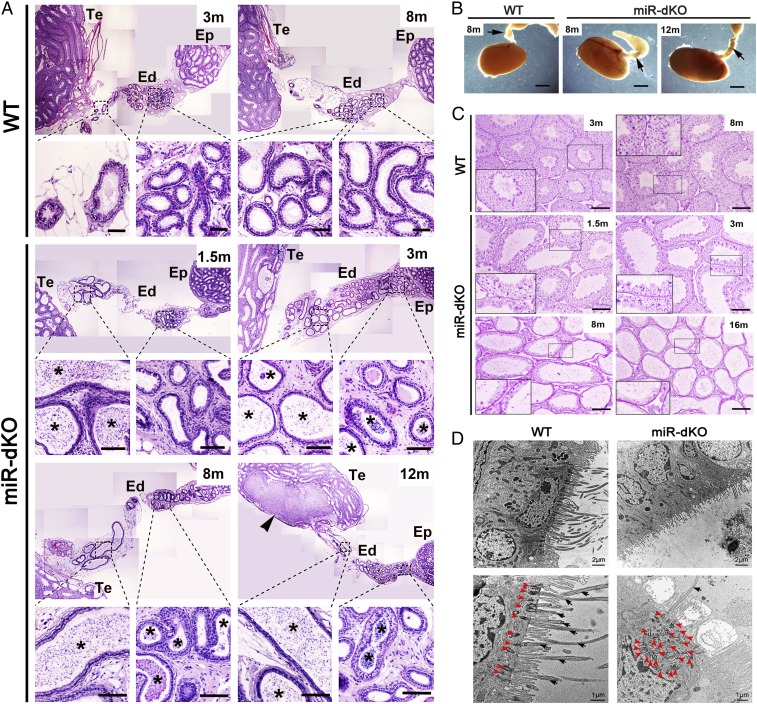
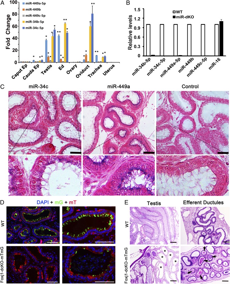
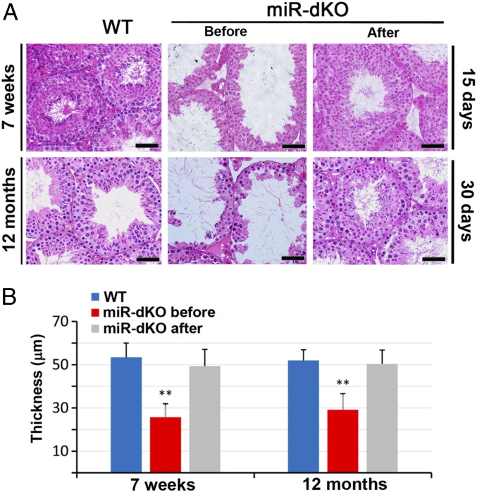
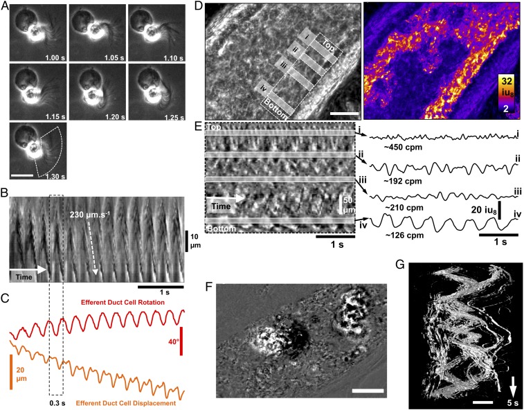
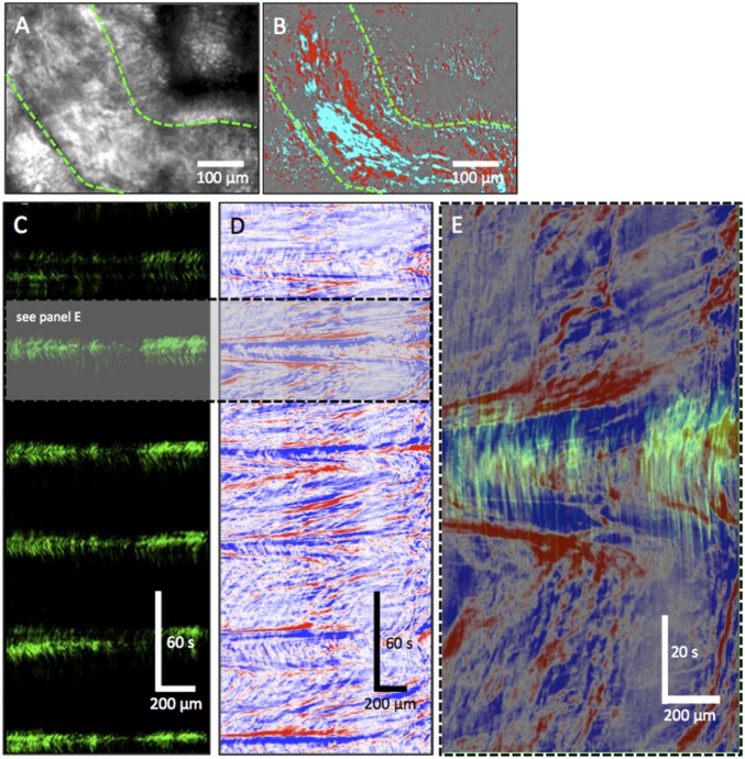
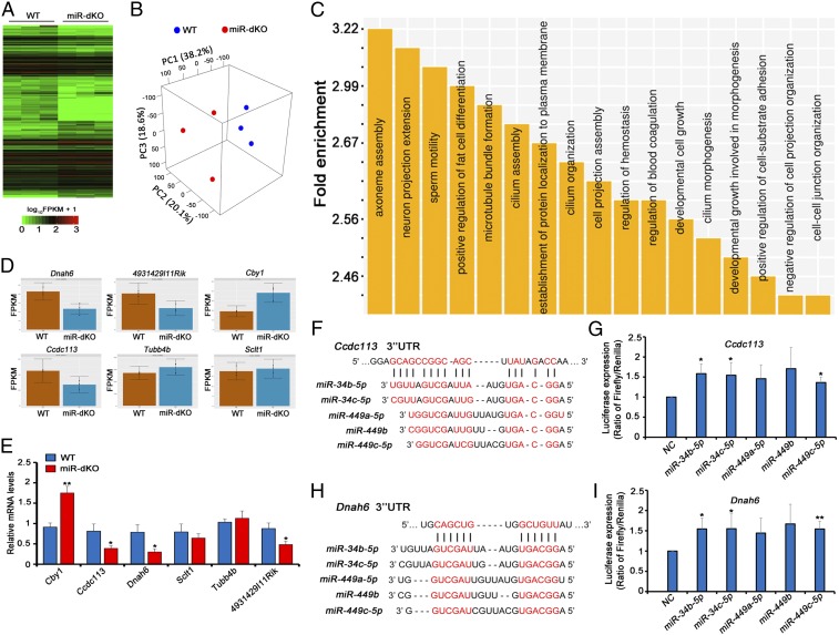
Cilia are cell-surface, microtubule-based organelles that project into extracellular space. Motile cilia are conserved throughout eukaryotes, and their beat induces the flow of fluid, relative to cell surfaces. In mammals, the coordinated beat of motile cilia provides highly specialized functions associated with the movement of luminal contents, as seen with metachronal waves transporting mucus in the respiratory tract. Motile cilia are also present in the male and female reproductive tracts. In the female, wave-like motions of oviductal cilia transport oocytes and embryos toward the uterus. A similar function has been assumed for motile cilia in efferent ductules of the male-i.e., to transport immotile sperm from rete testis into the epididymis. However, we report here that efferent ductal cilia in the male do not display a uniform wave-like beat to transport sperm solely in one direction, but rather exert a centripetal force on luminal fluids through whip-like beating with continual changes in direction, generating turbulence, which maintains immotile spermatozoa in suspension within the lumen. Genetic ablation of two miRNA clusters (miR-34b/c and -449a/b/c) led to failure in multiciliogenesis in murine efferent ductules due to dysregulation of numerous genes, and this mouse model allowed us to demonstrate that loss of efferent duct motile cilia causes sperm aggregation and agglutination, luminal obstruction, and sperm granulomas, which, in turn, induce back-pressure atrophy of the testis and ultimately male infertility.

Keywords: fluid resorption; male infertility; microRNA; multiciliogenesis; reproductive tract.


Conflict of interest statement

The authors declare no conflict of interest.

Figures

Fig. 1.
Fig. 1.
Spermatogenic disruptions, efferent ductal occlusion, and motile cilial defects in miR-dKO male mice. (A) H&E-stained paraffin sections showing that concentrated sperm/sperm agglutinations (asterisks) are present in the lumen of the EDs in miR-dKO males. While spermatozoa are rarely seen in the lumen of WT EDs in the proximal and conus segments, the miR-dKO EDs are filled with high concentrations of spermatozoa, initially only in the proximal segments at 1.5 mo of age, and then spread to the entire length thereafter (3, 8, and 12 mo of age). Sperm granuloma (arrowhead) is obvious in the rete testis of a 12-mo-old miR-dKO male mouse. Ep, epididymis; Te, testis. (Scale bars: 200 μm.) Note: Multiple panels of low-power images were assembled to view the entire EDs. (B) Transillumination patterns of the EDs in WT (8-mo-old) and miR-dKO (8- and 12-mo-old) mice. Arrows point to the EDs. WT ductules are smaller and nearly translucent, while miR-dKO ductules appear thicker and show more light absorption, indicative of ductules filled with stagnant spermatozoa. (Scale bars: 1 mm.) (C) H&E-stained paraffin sections showing that the miR-dKO testes display disrupted spermatogenesis characterized by reduced stratification of seminiferous epithelia and dilated lumens compared with WT testes. The severity of disruptions was increased with age (from 1.5 to 3, 8, and 16 mo). C, Insets show the digitally amplified subfields (framed). (Scale bars: 200 μm.) (D) Transmission electron microscopic images of the epithelium of WT and miR-dKO EDs. In miR-dKO ductules, fewer ciliated cells are observed, and, when present, only rare motile cilia are found extending into the lumen (black arrowheads). However, multiple disorganized centrioles (red arrowheads) are seen in the apical cytoplasm. Nonciliated cells appear normal with microvilli extending into the lumen. WT EDs display fully developed and well-aligned basal bodies along the surface of the ciliated cells, with numerous long motile cilia extending into the lumen.
Fig. 2.
Fig. 2.
miR-34b/c and -449a/b/c are abundantly expressed in the ciliated cells of EDs and mice with ciliated cell-specific knockout of the five miRNAs phenocopy the global double miRNA cluster knockout mice. (A) qPCR analyses on miR-34b/c and -449a/b/c expression levels in the caput and cauda epididymis (Ep), testis, EDs, ovary, oviduct, trachea, and uterus. U6 snRNA was used as an internal control, and the fold changes were calculated against levels in the caput epididymis. Data are presented as mean ± SEM (n = 3). *P < 0.05; **P < 0.01 (Student t test). (B) qPCR analyses on miR-34b/c and -449a/b/c expression levels in WT and miR-dKO EDs. miR-16 was used as an internal control. Data are presented as mean ± SEM (n = 3). (C) In situ hybridization results showing that both miR-34c and -449a are localized to the ciliated cells in the efferent ductal epithelium. Specific hybridization signals are blue. (Scale bars: 50 μm.) (D) Fluorescent microscopic images showing cross-sections of EDs in control (Foxj1-Cre;Rosa26mTmG+/Tg) and Foxj1-dcKO-mTmG (Foxj1-Cre;Rosa26mTmG+/Tg;miR-34b/c−/−;miR-449lox/lox) mice. Expression of membrane-bound EGFP (mG) is indicative of Cre-expressing ciliated cells. The mG-positive ciliated cells are far fewer in Foxj1-dcKO-mTmG EDs than in controls. (Scale bars: 100 μm.) (E) H&E-stained paraffin sections of testes and EDs in WT (Foxj1-Cre;Rosa26mTmG+/Tg) and Foxj1-dcKO-mTmG (Foxj1-Cre;Rosa26mTmG+/Tg;miR-34b/c−/−;miR-449lox/lox) mice. Ciliated cell-specific conditional knockout (Foxj1-dcKO) males appear to phenocopy the global knockout (miR-dKO) males because both display efferent ductal occlusion and sperm agglutination (arrows), as well as disrupted spermatogenesis due to dilated seminiferous tubules (asterisks). (Scale bars: 100 μm.)
Fig. 3.
Fig. 3.
Testicular histology showing full recovery of spermatogenesis in miR-dKO males after surgical relief of the back pressure. (A) Testes of 7-wk-old and 12-mo-old miR-dKO mice showed disrupted spermatogenesis characterized by reduced stratification of seminiferous epithelia and enlarged lumen indicative of back pressure due to fluid retention. By generating two tiny holes using a hooked surgical needle (diameter = 0.6 mm) in the rete testes, the back pressure was released, and full spermatogenesis, similar to that in WT testes, was observed 15 and 30 d after the procedure. (Scale bars: 40 μm.) (B) Quantitative analyses of the thickness of seminiferous epithelia in WT and miR-dKO testes before and after the surgical procedure to relieve the back pressure. Data are presented as mean ± SEM (n = 3). **P < 0.01 (Student t test).
Fig. 4.
Fig. 4.
Cilia beating patterns in isolated ciliated cells and intact EDs. (A) Image sequences showing the beating motion of cilia and the resulting rotation in a ciliated cell isolated from adult EDs. All frames are in the same scale. (Scale bar: 15 μm.) (B) An ST map (time in the x axis) showing changes in cilia opacity/translucency calculated perpendicularly to the overall arc of motion (see dashed region in A, Bottom). (C) Object displacement and rotation tracking of the isolated ciliated cell show the regularity of cilia motion and the propulsive effect on the cell. Efferent ductal cilia generate larger torque forces that rotate (±20° per beat) and displace the cell (±7–10 µm). (D) Image of the field of view (FOV) using power imaging of intact ED tube (Left) and SD of opacity/translucency demarcating those areas in which detectable cilia beating was observed (Right). (Scale bar: 100 μm.) (E, Left) An ST map (time in the x axis) taken from a region parallel to the ED wall showing distinct rhythmic frequencies (gray bars i–iv). (E, Right) Plotting the time courses of the changes in opacity/translucency reveals a wide variety of frequencies and amplitudes in adjacent regions [∼450 cpm (i) to ∼126 cpm (iv)]. (F) Differential image showing two clumps of materials (most likely cell clumps sloughed into the lumen during whole-mount preparation) within the EDs. (Scale bar: 100 μm.) (G) The 3D ST objects constructed from the two clumps of materials in F, showing the swirling motion applied to objects by the underlying movement of cilia. (Scale bar: 100 μm.)
Fig. 5.
Fig. 5.
Peristaltic contractions of the efferent ductal smooth muscle propel sperm to the epididymis. (A) Raw movie frame showing the efferent ductal wall (green dashed lines) and vessel lumen containing immotile sperm. (B) Differential imaging (∆t ± 5 s) was used to remove static background to better delineate wall motion and sperm. (C) ST map of wall motion (upper green edge) showing regular peristaltic contractions that occurred approximately every minute (interval 58.5 ± 4.7 s; o = 5) and lasted ∼20 s. (D) ST map of the movement of contents (sperm) within the lumen of the ED. Peristaltic contractions of the ED smooth muscle propelled sperm rapidly at velocities of 100–300 µm⋅s−1. In between peristaltic contractions, sperm movement was variable and slow (10–70 µm⋅s−1). (E) Overlay of ST maps of a peristaltic contraction (see gray region in C; green) and content movement (see gray region in D; red/blue) showing the rapid propulsion of sperm during a peristaltic contraction.
Fig. 6.
Fig. 6.
miR-34b/c and -449a/b/c control multiciliogenesis in the EDs by regulating the expression of key genes involved in ciliogenesis. (A) A hierarchical clustering heatmap showing 7,002 significantly dysregulated mRNAs in miR-dKO EDs. (B) A 3D-PCA plot of the first three principal components (PCs) showing the clusters between WT and miR-dKO samples. The first three PCs account for 76.9% of the total variance, and samples of WT are distinctly separated from those of miR-dKO. (C) GO term enrichment analyses of significantly dysregulated mRNAs in miR-dKO EDs. Outputs (biological processes) are sorted and plotted against fold enrichment. (D) Levels of six representative, ciliogenesis-associated genes predicted to be targeted by the five miRNAs (miR-34b/c and -449a/b/c) based on RNA-seq data. (E) qPCR validation of the levels of the six target genes, as shown in D, in WT and miR-dKO EDs. Gapdh was used as the internal control for normalization. Data are presented as mean ± SEM (n  =  3). *P < 0.05; **P < 0.01 (Student’s t test). (F and H) Schematics showing two predicted miR-34/449-binding sites in the 3′ UTRs of Ccdc113 (F) and Dnah6 (H). (G and I) Luciferase-based reporter assays showing miR-34b/c–miR-449c–dependent stabilization of Ccdc13 (G) and Dnah6 (I) in HEK293 cells. Data are presented as mean ± SEM (n = 3). *P < 0.05; **P < 0.01 (Student’s t test).

Comment in

References

    1. Satir P, Christensen ST. Structure and function of mammalian cilia. Histochem Cell Biol. 2008;129:687–693. - PMC - PubMed
    1. Braun DA, Hildebrandt F. Ciliopathies. Cold Spring Harb Perspect Biol. 2016;9:a028191. - PMC - PubMed
    1. Afzelius BA. Cilia-related diseases. J Pathol. 2004;204:470–477. - PMC - PubMed
    1. Wong LB, Miller IF, Yeates DB. Nature of the mammalian ciliary metachronal wave. J Appl Physiol (1985) 1993;75:458–467. - PubMed
    1. O’Callaghan C, Sikand K, Chilvers MA. Analysis of ependymal ciliary beat pattern and beat frequency using high speed imaging: Comparison with the photomultiplier and photodiode methods. Cilia. 2012;1:8. - PMC - PubMed

Publication types

LinkOut - more resources