

Blocking CXCLs-CXCR2 axis in tumor-stromal interactions contributes to survival in a mouse model of pancreatic ductal adenocarcinoma through reduced cell invasion/migration and a shift of immune-inflammatory microenvironment
- PMID: 30659170
- PMCID: PMC6338726
- DOI: 10.1038/s41389-018-0117-8
Blocking CXCLs-CXCR2 axis in tumor-stromal interactions contributes to survival in a mouse model of pancreatic ductal adenocarcinoma through reduced cell invasion/migration and a shift of immune-inflammatory microenvironment
Abstract
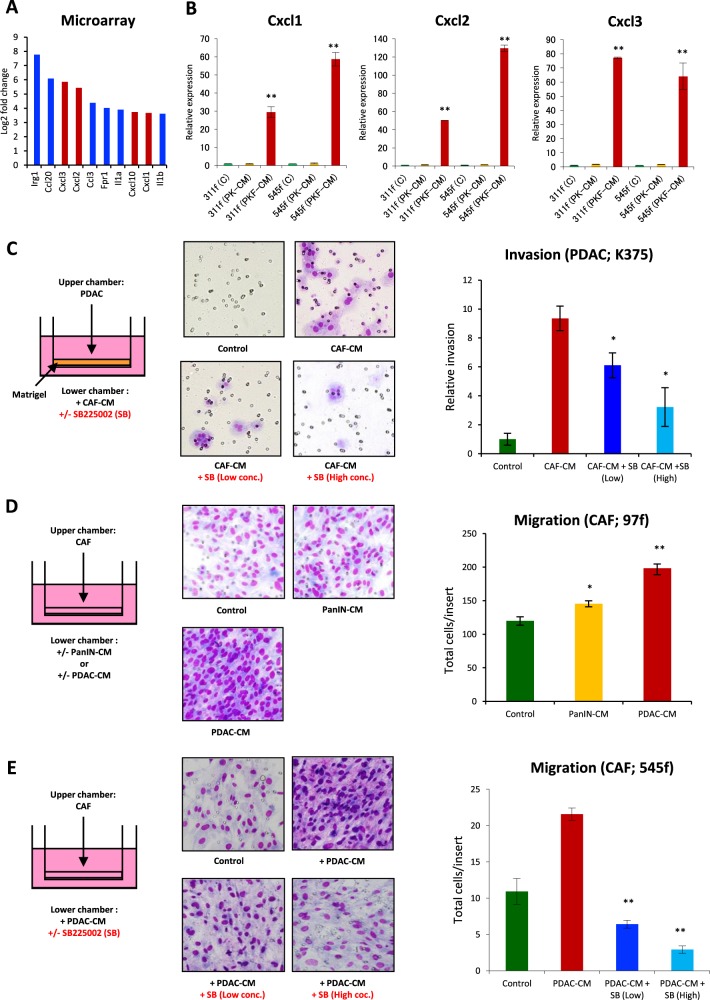
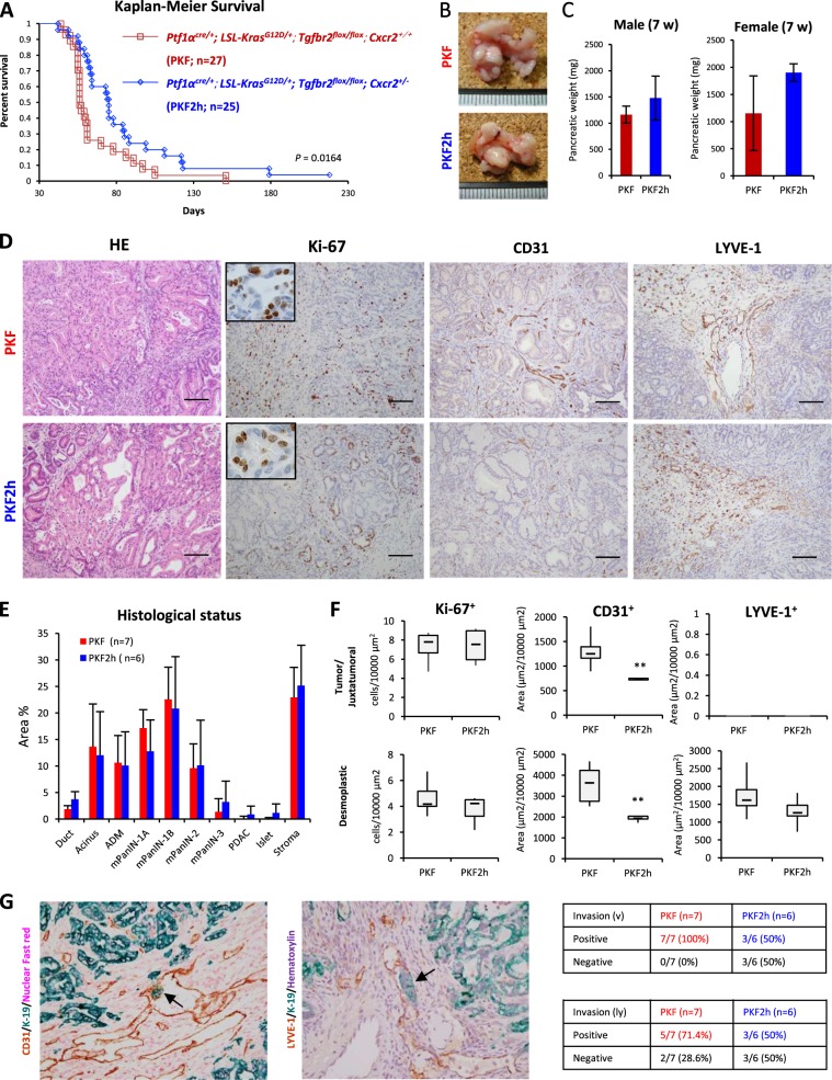
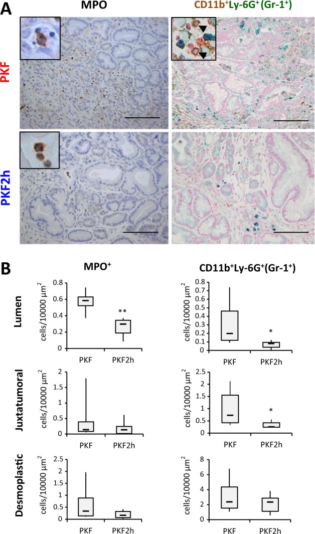
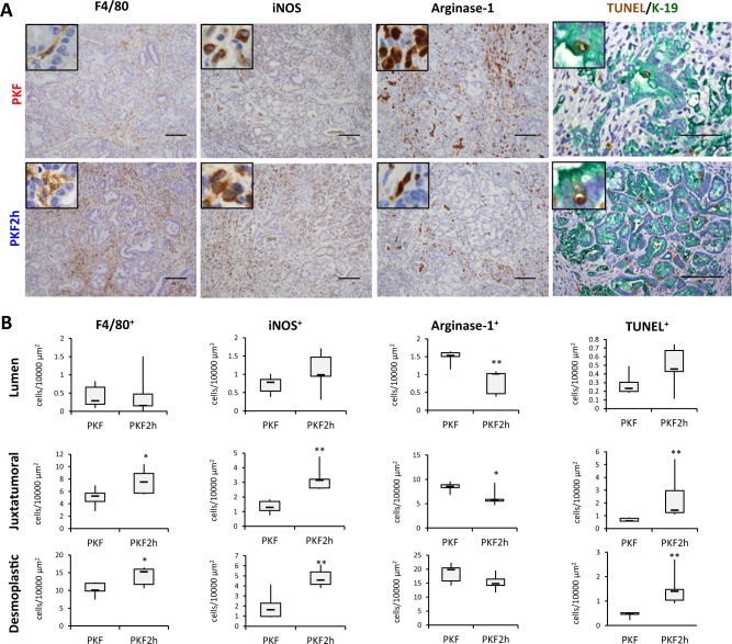
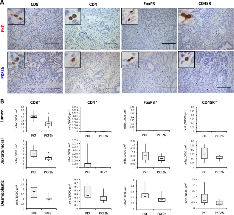
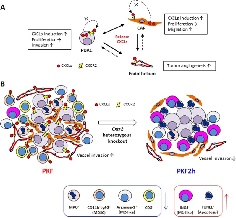
Pancreatic ductal adenocarcinoma (PDAC) is characterized by dense stromal reaction (desmoplasia). We have previously reported that mice with conditional KrasG12D mutation and knockout of TGF-β receptor type II (Tgfbr2), PKF mice, develop PDAC with desmoplasia modulated by CXC chemokines that are produced by PDAC cells through tumor-stromal interaction. In this study, we further discovered that PDAC and cancer-associated fibroblast (CAF) accelerated each other's invasion and migration through the CXC chemokines-receptor (CXCLs-CXCR2) axis. Heterozygous knockout of Cxcr2 in PKF mice (PKF2h mice) prolonged survival and inhibited both tumor angiogenesis and PDAC microinvasion. Infiltration of neutrophils, myeloid-derived suppressor cells (MDSCs), and arginase-1+ M2-like tumor-associated macrophages (TAMs) significantly decreased in the tumors of PKF2h mice, whereas inducible nitric oxide synthase (iNOS)+ M1-like TAMs and apoptotic tumor cells markedly increased, which indicated that blockade of the CXCLs-CXCR2 axis resulted in a shift of immune-inflammatory microenvironment. These results suggest that blocking of the CXCLs-CXCR2 axis in tumor-stromal interactions could be a therapeutic approach against PDAC progression.
Conflict of interest statement
The authors declare that they have no conflict of interest.
Figures
Similar articles
-
Inhibiting Cxcr2 disrupts tumor-stromal interactions and improves survival in a mouse model of pancreatic ductal adenocarcinoma.J Clin Invest. 2011 Oct;121(10):4106-17. doi: 10.1172/JCI42754. Epub 2011 Sep 19. J Clin Invest. 2011. PMID: 21926469 Free PMC article.
-
Modulating the CXCR2 Signaling Axis Using Engineered Chemokine Fusion Proteins to Disrupt Myeloid Cell Infiltration in Pancreatic Cancer.Biomolecules. 2025 Apr 30;15(5):645. doi: 10.3390/biom15050645. Biomolecules. 2025. PMID: 40427538 Free PMC article.
-
Potential roles and targeted therapy of the CXCLs/CXCR2 axis in cancer and inflammatory diseases.Biochim Biophys Acta Rev Cancer. 2019 Apr;1871(2):289-312. doi: 10.1016/j.bbcan.2019.01.005. Epub 2019 Jan 29. Biochim Biophys Acta Rev Cancer. 2019. PMID: 30703432 Review.
-
Targeting both tumour-associated CXCR2+ neutrophils and CCR2+ macrophages disrupts myeloid recruitment and improves chemotherapeutic responses in pancreatic ductal adenocarcinoma.Gut. 2018 Jun;67(6):1112-1123. doi: 10.1136/gutjnl-2017-313738. Epub 2017 Dec 1. Gut. 2018. PMID: 29196437 Free PMC article.
-
Desmoplasia in pancreatic ductal adenocarcinoma: insight into pathological function and therapeutic potential.Genes Cancer. 2018 Mar;9(3-4):78-86. doi: 10.18632/genesandcancer.171. Genes Cancer. 2018. PMID: 30108679 Free PMC article. Review.
Cited by
-
CXCR1: A Cancer Stem Cell Marker and Therapeutic Target in Solid Tumors.Biomedicines. 2023 Feb 16;11(2):576. doi: 10.3390/biomedicines11020576. Biomedicines. 2023. PMID: 36831112 Free PMC article. Review.
-
CXCL3 Signaling in the Tumor Microenvironment.Adv Exp Med Biol. 2021;1302:15-24. doi: 10.1007/978-3-030-62658-7_2. Adv Exp Med Biol. 2021. PMID: 34286438 Review.
-
The tumor microenvironment in pancreatic ductal adenocarcinoma: current perspectives and future directions.Cancer Metastasis Rev. 2021 Sep;40(3):675-689. doi: 10.1007/s10555-021-09988-w. Cancer Metastasis Rev. 2021. PMID: 34591240 Review.
-
Next-Generation Immunotherapies to Improve Anticancer Immunity.Front Pharmacol. 2021 Jan 11;11:566401. doi: 10.3389/fphar.2020.566401. eCollection 2020. Front Pharmacol. 2021. PMID: 33505304 Free PMC article. Review.
-
Murine Macrophages Modulate Their Inflammatory Profile in Response to Gas Plasma-Inactivated Pancreatic Cancer Cells.Cancers (Basel). 2021 May 21;13(11):2525. doi: 10.3390/cancers13112525. Cancers (Basel). 2021. PMID: 34064000 Free PMC article.
References
-
- Kato, K. Vital Statistics in Japan. Director-General for statistics and Information Policy, Ministry of Health, Labour and Welfare, Tokyo, pp. 18–19 (2017).
LinkOut - more resources
Full Text Sources
Research Materials
Miscellaneous
