. 2019 Jan 18;10(1):338.
doi: 10.1038/s41467-018-08187-6.

Malonylation of GAPDH is an inflammatory signal in macrophages

Affiliations

Malonylation of GAPDH is an inflammatory signal in macrophages

Silvia Galván-Peña et al. Nat Commun. .

Abstract

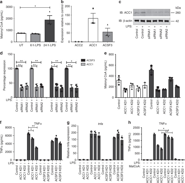
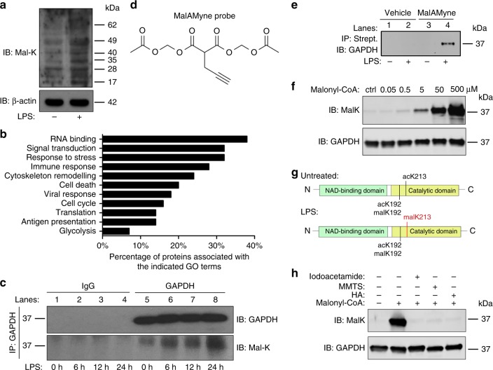
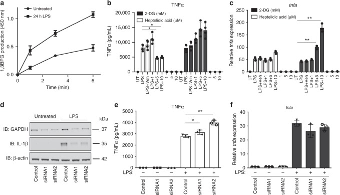
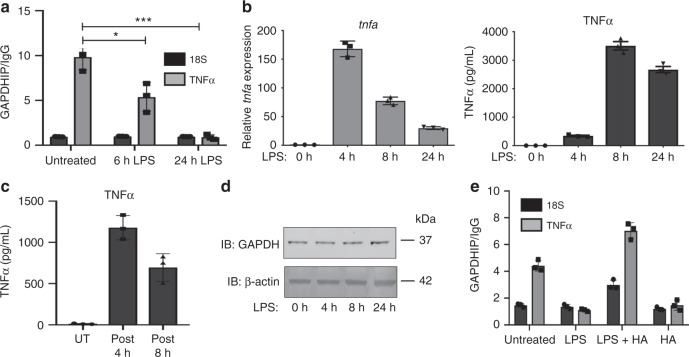
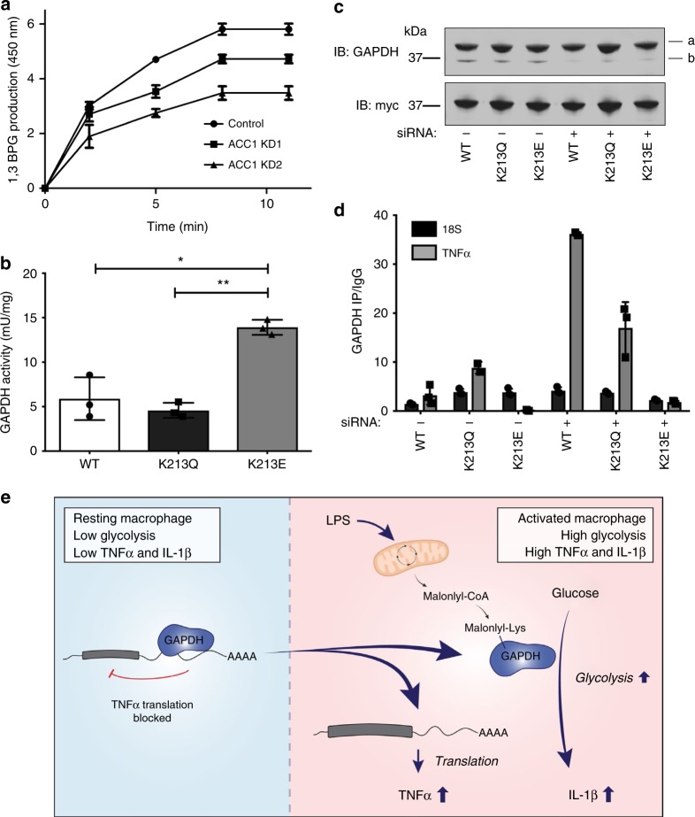
Macrophages undergo metabolic changes during activation that are coupled to functional responses. The gram negative bacterial product lipopolysaccharide (LPS) is especially potent at driving metabolic reprogramming, enhancing glycolysis and altering the Krebs cycle. Here we describe a role for the citrate-derived metabolite malonyl-CoA in the effect of LPS in macrophages. Malonylation of a wide variety of proteins occurs in response to LPS. We focused on one of these, glyceraldehyde-3-phosphate dehydrogenase (GAPDH). In resting macrophages, GAPDH binds to and suppresses translation of several inflammatory mRNAs, including that encoding TNFα. Upon LPS stimulation, GAPDH undergoes malonylation on lysine 213, leading to its dissociation from TNFα mRNA, promoting translation. We therefore identify for the first time malonylation as a signal, regulating GAPDH mRNA binding to promote inflammation.


Conflict of interest statement

The authors declare no competing interests.

Figures

Fig. 1
Fig. 1
Activation of macrophages increases malonyl-CoA levels and is needed for pro-inflammatory cytokine production. a Malonyl-CoA levels measured in untreated and LPS-treated (100 ng/mL) BMDM lysates using a malonyl-CoA ELISA. Mean + SEM, n = 3. b ACC1, ACC2 and ACSF3 mRNA expression measured via qPCR in BMDMs. Mean + SEM, n = 3. c ACC1 siRNA (48 h, 10 nM) knockdown (KD) measured by western blotting in untreated and LPS-treated (100 ng/mL, 24 h) BMDMs. β-actin used as a control. Representative of three independent experiments. d ACC1 and ACSF3 siRNA (48 h, 10 nM) KD levels relative to control siRNA, measured via qPCR in untreated and LPS-treated (100 ng/mL, 24 h) BMDMs. Mean + SEM, n = 4. e Malonyl-CoA levels measured in ACC1 and ACSF3 KD BMDM lysates using a malonyl-CoA ELISA. Mean + SD, representative of four independent experiments. f TNFα protein measured by ELISA (mean + SEM, n = 3) and g TNFα mRNA expression in 100 ng/mL LPS-treated (100 ng/mL, 6 h) ACC1 and ACSF3 KD BMDMs (mean + SD, representative of three independent experiments). h TNFα protein measured by ELISA in ACC1 KD BMDMs pre-treated with malonylCoA (1 mM, 2 h) followed by LPS (100 ng/mL, 6 h) (mean + SD, representative of three independent experiments). Unpaired t-test, *P< 0.05; **P< 0.005
Fig. 2
Fig. 2
Activation of macrophages with LPS increases protein malonylation, with GAPDH as a substrate. a Western blot analysis of lysine malonylation (mal-K) in lysates from BMDMs treated with LPS (100 ng/mL) for 24 h. b Most enriched functions associated with LPS-induced malonylated proteins (FDR < 0.05). c Immunoprecipitated GAPDH from untreated and LPS-treated (100 ng/mL, 6, 12 and 24 h) BMDMs and samples probed with an anti-malK antibody (lower panel). GAPDH expression in the immunoprecipitated (upper panel) samples was also examined. d Chemical formula of the MalAMyne probe. e Untreated and LPS-treated (100 ng/mL, 24 h) BMDMs were labelled with MalAMyne (10 µM) or vehicle control. Copper-catalysed click chemistry was performed on the lysates using biotin, followed by immunoprecipitation using streptavidin (strept.). Samples were probed for GAPDH via western blotting. f Purified GAPDH (100 µg/mL) was incubated in the presence of TCEP and malonyl-CoA (or buffer as control) for 1 h at 37 °C, pH 7.5. K-malonylation was assessed by western blotting. g Identification of malonylated sites in immunoprecipitated trypsin-digested GAPDH peptides from untreated and LPS-treated BMDMs (106 cells). Peptides were analysed via LC–MS. h Purified GAPDH was preincubated with TCEP and 25 mM iodoacetamide, 80 mM methyl methanethiosulfonate (MMTS), 5 μM heptelidic acid (HA) or buffer for 30 min at 37 °C, followed by 500 μM malonyl-CoA. K-malonylation was assessed by western blotting. All data shown are representative of three independent experiments
Fig. 3
Fig. 3
GAPDH controls TNFα production. a Untreated, 6 and 24 h LPS-treated (100 ng/mL) BMDM lysates were assayed for GAPDH enzymatic activity by monitoring product production (1,3 bisphosphoglycerate, 1,3BPG) over time. Representative of three independent experiments. b TNFα protein measured by MSD (mean + SEM, n = 4) and c TNFα mRNA expression in 100 ng/mL LPS-treated BMDMs (6 h), pre-treated with HA or 2-DG. d GAPDH siRNA (72 h, 10 nM) KD in BMDMs, measured by western blotting. Pro-IL1β levels were also measured, and β-actin used as a control. Representative of three independent experiments. e TNFα measured in GAPDH KD BMDMs, treated with LPS (24 h, 100 ng/mL, mean + SEM, n = 3). f TNFα mRNA expression measured by qPCR in LPS-treated GAPDH KD BMDMs. Unpaired t-test, *p < 0.05; **p < 0.01
Fig. 4
Fig. 4
GAPDH regulates TNFα post-transcriptional regulation. a GAPDH was immunoprecipitated from untreated and LPS-treated (6 and 24 h, 100 ng/mL) BMDMs, and TNFα mRNA presence in IP relative to IgG control assessed via qPCR. (mean + SEM, n = 3). b TNFα mRNA expression measured by qPCR and TNFα protein measured by ELISA in BMDMs treated with LPS (100 ng/mL) over time. c TNFα protein measured by ELISA in BMDMs treated with LPS (100 ng/mL). Cells were washed post 4 h and post 8 h LPS treatment, and supernatants replaced (without LPS) and harvested 20 and 16 h later, respectively (mean + SEM, n = 3). d GAPDH expression analysed by western blotting in LPS-treated BMDMs. e LPS-treated (100 ng/mL, 24 h) BMDMs were pre-treated with HA (10 µM) and GAPDH immunoprecipitated. Bound TNFα mRNA was assessed by qPCR. Data shown are representative of three independent experiments. Unpaired t-test, *p < 0.05; ***p < 0.01
Fig. 5
Fig. 5
K213 malonylation regulates GAPDH enzymatic activity and RNA-binding. a GAPDH enzymatic activity measured in lysates from ACC1 and ACSF3 KD BMDMs (LPS treated, 100 ng/mL, 24 h; mean + SD). b WT, K213Q and K213E GAPDH mutants were overexpressed in HEK293T, affinity purified, and the enzymatic activity measured (mean + SEM, n = 3). c GAPDH was knockdown using siRNA (48 h, 10 nM) followed by overexpression of WT, K213Q and K213E GAPDH mutants and expression assessed via western blotting (a, overexpressed GAPDH; b, endogenous GAPDH). d GAPDH mutants were immunoprecipitated following 48 h overexpression and fixing in HEK293T cells. Bound TNFα mRNA in the IP relative to IgG was assessed via qPCR. Mean + SD, representative of three independent experiments. Unpaired t-test, *p < 0.05; **p < 0.01. e In resting macrophages, GAPDH binds to the 3′-UTR of various pro-inflammatory mediators, such as TNFα, and prevents their translation. Following activation of macrophages with LPS, there is an accumulation of citrate that can be converted into malonyl-CoA in an ACC1-dependent manner. Malonyl-CoA can then in turn mediate an increase in lysine malonylation. One of the substrates of this modification is GAPDH, which after undergoing malonylation, releases the bound RNAs which can now be translated. At the same time, GAPDH enzymatic activity increases, which allows for an increased glycolytic flux, required for the activated macrophage to carry out its pro-inflammatory functions

References

    1. Karve TM, Cheema AK. Small changes huge impact: the role of protein posttranslational modifications in cellular homeostasis and disease. J. Amino Acids. 2011;2011:207691. doi: 10.4061/2011/207691. - DOI - PMC - PubMed
    1. Peng C, et al. The first identification of lysine malonylation substrates and its regulatory enzyme. Mol. Cell. Proteom. 2011;10:M111 012658. doi: 10.1074/mcp.M111.012658. - DOI - PMC - PubMed
    1. Xie Z, et al. Lysine succinylation and lysine malonylation in histones. Mol. Cell. Proteom. 2012;11:100–107. doi: 10.1074/mcp.M111.015875. - DOI - PMC - PubMed
    1. Bowman CE, et al. The mammalian malonyl-CoA synthetase ACSF3 is required for mitochondrial protein malonylation and metabolic efficiency. Cell Chem. Biol. 2017;24:673–684 e674. doi: 10.1016/j.chembiol.2017.04.009. - DOI - PMC - PubMed
    1. Colak G, et al. Proteomic and biochemical studies of lysine malonylation suggest its malonic aciduria-associated regulatory role in mitochondrial function and fatty acid oxidation. Mol. Cell. Proteom. 2015;14:3056–3071. doi: 10.1074/mcp.M115.048850. - DOI - PMC - PubMed

Publication types

MeSH terms