. 2019 Jan 18;10(1):314.
doi: 10.1038/s41467-018-08282-8.

Noninvasive optical activation of Flp recombinase for genetic manipulation in deep mouse brain regions

Affiliations

Noninvasive optical activation of Flp recombinase for genetic manipulation in deep mouse brain regions

Hyunjin Jung et al. Nat Commun. .

Abstract

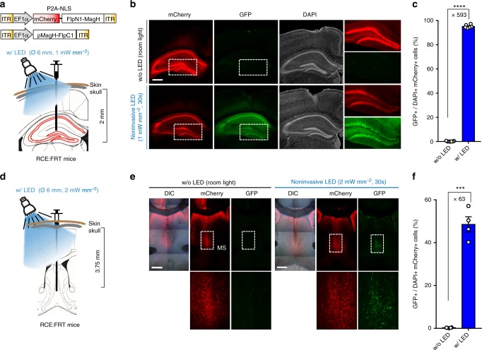
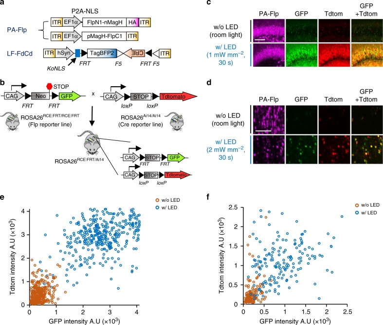
Spatiotemporal control of gene expression or labeling is a valuable strategy for identifying functions of genes within complex neural circuits. Here, we develop a highly light-sensitive and efficient photoactivatable Flp recombinase (PA-Flp) that is suitable for genetic manipulation in vivo. The highly light-sensitive property of PA-Flp is ideal for activation in deep mouse brain regions by illumination with a noninvasive light-emitting diode. In addition, PA-Flp can be extended to the Cre-lox system through a viral vector as Flp-dependent Cre expression platform, thereby activating both Flp and Cre. Finally, we demonstrate that PA-Flp-dependent, Cre-mediated Cav3.1 silencing in the medial septum increases object-exploration behavior in mice. Thus, PA-Flp is a noninvasive, highly efficient, and easy-to-use optogenetic module that offers a side-effect-free and expandable genetic manipulation tool for neuroscience research.


Conflict of interest statement

The authors declare no competing interests.

Figures

Fig. 1
Fig. 1
Development of PA-Flp. a Schematic depicting PA-Flp reconstitution and activation upon blue light illumination, and detection of GFP signals by PA-Flp-mediated deletion of a stop cassette in a Frt-floxed construct. b PA-Flp and fDIO-YFP (Flp reporter) expression plasmids were electroporated into an embryonic mouse brain (E15), with (w/) subsequent noninvasive light stimulation (0.5 mW mm−2, blue fluorescent gun) at postnatal day 1–2 (P1–2) or maintained in dark conditions. Pup brains were harvested at P3-4. c AAV-EF1a-PA-Flp and AAV-EF1a-fDIO-YFP were co-infected into the hippocampus of 8-wk-old mice, with 30 min light (0.4 mW mm−2, 20 Hz, 20% duty cycle) or without (w/o) light stimulation 2 wks after infection and sacrificed 1 wk after light stimulation. A blue line indicates laser light path through implanted optic fiber. b, c Measurement of GFP positive cells among both mCherry and iRFP positive (GFP+/mCh+iRFP+) cells or among both DAPI and mCherry positive (GFP+/DAPI+mCh+) cells in 4–8 coronal slices at each group. Scale bar: 100 μm. Data represent means ± s.e.m. (n = 2 mice/group; ***P< 0.0001, two-tailed Student’s t-test). d, e Schematic depicting AAV infection in the mouse hippocampus and local light stimulation via implanted optic fiber (d), and AAV targeting sites (green) and optic fiber implantation sites (red) (e), in coronal sections of hippocampus. f Left: representative images showing local labeling of GFP signals from RCE:FRT mice at different depths along the rostral-caudal (RC) axis. Yellow arrowhead indicates the medial-lateral (ML) 1.35 coordinate. Right: sites (i)–(iv) at left marked in white-dashed squares are shown in higher-magnification views in the correspondingly labeled rows. Sites (i), (ii), and (iv) indicate the same ML coordinate along the RC axis; (iii) indicates a distal site with a different ML coordinate on the same coronal section of bregma (−1.88 mm). Scale bar: 100 μm. g, h Analysis of percentage (g) and intensity (h) of GFP signals along the ML axis at the same RC coordinate (bregma −1.88 mm) or RC axis on the same ML coordinate (ML 1.35). Measurement performed in each coronal slice. Data represent means ± s.e.m. (n = 1 mouse)
Fig. 2
Fig. 2
Noninvasive LED illumination activates PA-Flp in deep brain structures down to the hippocampus and MS. a, d Schematic depicting AAV-EF1a-PA-Flp targeting in the hippocampus (a) or MS (d) followed by LED illumination. b, e Representative images of mCherry (PA-Flp) and GFP (Flp reporter) signals from RCE:FRT mice (8–12-wk-old), with (w/) and without (w/o) LED illumination. Two wks after infection, light was illuminated noninvasively with white LED at an intensity of 1 mW mm−2 (b) or 2 mW mm−2 (e) for 30 s through the intact skull and skin. Mice were maintained under room light as described in Methods. All mice were sacrificed 3 wks after infection. Scale bar: 500 μm. c, f GFP positive cells among both DAPI and mCherry positive (GFP+/DAPI+mCh+) cells were measured in the hippocampus (c) and MS (f) region of 4–7 coronal slices, as shown in b and e, respectively. Data represent means ± s.e.m. (c, n = 4 mice/group, ****P< 1 × 10⁻¹⁰; f, n = 2 mice/group, ***P< 0.0001; two-tailed Student’s t-test)
Fig. 3
Fig. 3
Verification of PA-Flp-dependent Cre (PA-FdCre) system in ROSA26RCE:FRT/Ai14 mouse line. a Schematic depicting AAVs carrying PA-Flp and Leak Free Flp-dependent Cre driver (LF-FdCd) as PA-Flp dependent Cre (PA-FdCre) systems. b Schematic depicting the generation of ROSA26RCE:FRT/Ai14 mice. Flp reporter (GFP) and Cre reporter (TdTomato) alleles are located on the ROSA26 cassette in ROSA26RCE:FRT/Ai14 mice. c, d Light-induced dual Flp and Cre activation in the hippocampal DG (c) or MS (d) of ROSA26RCE:FRT/Ai14 mice (8–12-wk-old). Scale bar: 100 μm. e, f Correlation between GFP and TdTomato fluorescence intensity in the hippocampal DG (e) or MS (f) among PA-Flp–positive cells (magenta) in with (w/) LED and without (w/o) LED groups. A total of 200–500 cells were counted from each w/ LED and w/o LED group (n = 4 mice/group)
Fig. 4
Fig. 4
PA-FdCre–mediated Cav3.1 knockdown in the MS increases object exploration. a Injection of a mixture of AAVs expressing PA-Flp, LF-FdCd and flox-shCav3.1 (or flox-shControl) into the MS of a 8-wk-old mice brain, with (w/) or without (w/o) subsequent noninvasive white LED (2 mW mm−2) illumination 2 wk after infection. b Schematic depicting object-exploration behavior in PA-FdCre–mediated, MS-specific, Cav3.1-knockdown mice. c Immunohistochemical detection of Cav3.1 in the MS following co-infection with PA-Flp-, LF-FdCd- and flox-shCav3.1-expressing AAVs. PA-Flp (mCh) and flox-shCav3.1 (GFP) fluorescence, Cav3.1 immunofluorescence (violet), and merged images are presented. Arrowheads indicate Cav3.1-positive cells. Arrows indicate PA-Flp/flox-shCav3.1–positive, Cav3.1-negative cells. Scale bar: 50 μm. d Cumulative traces of representative navigation pathways of shCav3.1 with LED and shCav3.1 without LED mice during object-exploration behavior. e Increased object-exploration time for shCav3.1 w/ LED mice (n = 9/group) compared with shCav3.1 w/o LED mice (n = 8; P = 0.003, two-way repeated measures ANOVA; P = 0.004, Bonferroni post-hoc test), shControl w/ LED mice (n = 5; P = 0.003, two-way repeated measures ANOVA; P = 0.028, Bonferroni post-hoc test), and shControl w/o LED (n = 5, P = 0.003, two-way repeated measures ANOVA; P = 0.009, Bonferroni post-hoc test). f Increased total object-exploration time for shCav3.1 w/ LED mice (n = 9) compared with that for shCav3.1 w/o LED mice (n = 8; P = 0.003, one-way ANOVA; P = 0.004, Bonferroni post-hoc test), shControl w/ LED mice (n = 5; P = 0.003, one-way ANOVA; P = 0.029, Bonferroni post-hoc test), and shControl w/o LED mice (n = 5; P = 0.003, one-way ANOVA; P = 0.008, Bonferroni post-hoc test). g Locomotor activity of shCav3.1 w/ LED (n = 9), shCav3.1 w/o LED (n = 8), shControl w/ LED mice (n = 5), and shControl w/o LED (n = 5) mice in the experimental cage during the 60-min habituation period of the object-exploration task (P = 0.612, one-way ANOVA). eg Data represent means ± s.e.m. (*P < 0.05, **P < 0.01)

Comment in

Similar articles

Cited by

References

    1. Wehr M, et al. Transgenic silencing of neurons in the mammalian brain by expression of the allatostatin receptor (AlstR) J. Neurophysiol. 2009;102:2554–2562. doi: 10.1152/jn.00480.2009. - DOI - PMC - PubMed
    1. Metzger D, Clifford J, Chiba H, Chambon P. Conditional site-specific recombination in mammalian cells using a ligand-dependent chimeric Cre recombinase. Proc. Natl Acad. Sci. USA. 1995;92:6991–6995. doi: 10.1073/pnas.92.15.6991. - DOI - PMC - PubMed
    1. Szobota S, et al. Remote control of neuronal activity with a light-gated glutamate receptor. Neuron. 2007;54:535–545. doi: 10.1016/j.neuron.2007.05.010. - DOI - PubMed
    1. Boyden ES, Zhang F, Bamberg E, Nagel G, Deisseroth K. Millisecond-timescale, genetically targeted optical control of neural activity. Nat. Neurosci. 2005;8:1263–1268. doi: 10.1038/nn1525. - DOI - PubMed
    1. Dong S, Rogan SC, Roth BL. Directed molecular evolution of DREADDs: a generic approach to creating next-generation RASSLs. Nat. Protoc. 2010;5:561–573. doi: 10.1038/nprot.2009.239. - DOI - PubMed

Publication types