

Hemispheric Regional Based Analysis of Diffusion Tensor Imaging and Diffusion Tensor Tractography in Patients with Temporal Lobe Epilepsy and Correlation with Patient outcomes
- PMID: 30659215
- PMCID: PMC6338779
- DOI: 10.1038/s41598-018-36818-x
Hemispheric Regional Based Analysis of Diffusion Tensor Imaging and Diffusion Tensor Tractography in Patients with Temporal Lobe Epilepsy and Correlation with Patient outcomes
Abstract
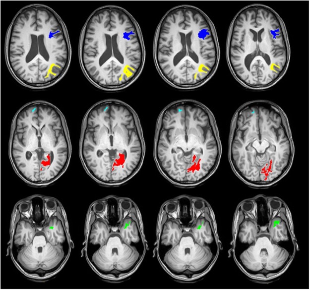
Imaging in the field of epilepsy surgery remains an essential tool in terms of its ability to identify regions where the seizure focus might present as a resectable area. However, in many instances, an obvious structural abnormality is not visualized. This has created the opportunity for new approaches and imaging innovation in the field of epilepsy, such as with Diffusion Tensor Imaging (DTI) and Diffusion Tensor Tractography (DTT). In this study, we aim to evaluate the use of DTI and DTT as a predictive model in the field of epilepsy, specifically Temporal Lobe Epilepsy (TLE), and correlate their clinical significance with respect to postsurgical outcomes. A hemispheric based analysis was used to compare the tract density, as well as DTI indices of the specific regions of interest from the pathologic hemisphere to the healthy hemisphere in TLE patients. A total of 22 patients with TLE (12 males, 10 females, 22-57 age range) underwent either a craniotomy, Anterior Temporal Lobectomy (ATL), or a less invasive method of Selective Laser Amygdalohippocampectomy (SLAH) and were imaged using 3.0 T Philips Achieva MR scanner. Of the participants, 12 underwent SLAH while 10 underwent ATL. The study was approved by the institutional review board of Thomas Jefferson University Hospital. Informed consent was obtained from all patients. All patients had a diagnosis of TLE according to standard clinical criteria. DTI images were acquired axially in the same anatomical location prescribed for the T1-weighted images. The raw data set consisting of diffusion volumes were first corrected for eddy current distortions and motion artifacts. Various DTI indices such as Fractional Anisotropy (FA), Mean Diffusivity (MD), Radial Diffusivity (RD) and Axial Diffusivity (AD) were estimated and co-registered to the brain parcellation map obtained by freesurfer. 16 consolidated cortical and subcortical regions were selected as regions of interest (ROIs) by a functional neurosurgeon and DTI values for each ROI were calculated and compared with the corresponding ROI in the opposite hemisphere. Also, track density imaging (TDI) of 68 white matter parcels were generated using fiber orientation distribution (FOD) based deterministic fiber tracking and compared with contralateral side of the brain in each epileptic group: left mesial temporal sclerosis (LMTS) and right MTS (RMTS)). In patients with LMTS, MD and RD values of the left hippocampus decreased significantly using two-tailed t-test (p = 0.03 and p = 0.01 respectively) compared to the right hippocampus. Also, RD showed a marginally significant decrease in left amygdala (p = 0.05). DTT analysis in LMTS shows a marginally significant decrease in the left white matter supramarginal parcel (p = 0.05). In patients with RMTS, FA showed a significant decrease in the ipsilateral mesial temporal lobe (p = 0.02), parahippocampal area (p = 0.03) and thalamus (p = 0.006). RD showed a marginally significant increase in the ipsilateral hippocampus (p = 0.05) and a significant increase in the ipsilateral parahippocampal area (p = 0.03). Also, tract density of the ipsilateral white matter inferior parietal parcel showed a marginally significant increase compared to the contralateral side (p = 0.05). With respect to postsurgical outcomes, we found an association between residual seizures and tract density in five white matter segments including ipsilateral lingual (p = 0.04), ipsilateral temporal pole (p = 0.007), ipsilateral pars opercularis (p = 0.03), ipsilateral inferior parietal (p = 0.04) and contralateral frontal pole (p = 0.04). These results may have the potential to be developed into imaging prognostic markers of postoperative outcomes and provide new insights for why some patients with TLE continue to experience postoperative seizures if pathological/clinical correlates are further confirmed.
Conflict of interest statement
The authors declare no competing interests.
Figures

Similar articles
-
Investigation of microstructural abnormalities in white and gray matter around hippocampus with diffusion tensor imaging (DTI) in temporal lobe epilepsy (TLE).Epilepsy Behav. 2018 Jun;83:44-49. doi: 10.1016/j.yebeh.2017.12.002. Epub 2018 Apr 10. Epilepsy Behav. 2018. PMID: 29653337
-
Group-specific regional white matter abnormality revealed in diffusion tensor imaging of medial temporal lobe epilepsy without hippocampal sclerosis.Epilepsia. 2010 Apr;51(4):529-35. doi: 10.1111/j.1528-1167.2009.02327.x. Epub 2009 Oct 8. Epilepsia. 2010. PMID: 19817819
-
White matter abnormalities associate with type and localization of focal epileptogenic lesions.Epilepsia. 2015 Jan;56(1):125-32. doi: 10.1111/epi.12871. Epub 2014 Dec 26. Epilepsia. 2015. PMID: 25545559
-
White matter in temporal lobe epilepsy: clinico-pathological correlates of water diffusion abnormalities.Quant Imaging Med Surg. 2015 Apr;5(2):264-78. doi: 10.3978/j.issn.2223-4292.2015.02.06. Quant Imaging Med Surg. 2015. PMID: 25853084 Free PMC article. Review.
-
Diffusion tensor imaging in Alzheimer's disease and mild cognitive impairment.Behav Neurol. 2009;21(1):39-49. doi: 10.3233/BEN-2009-0234. Behav Neurol. 2009. PMID: 19847044 Free PMC article. Review.
Cited by
-
Local and distant cortical responses to single pulse intracranial stimulation in the human brain are differentially modulated by specific stimulation parameters.Brain Stimul. 2022 Mar-Apr;15(2):491-508. doi: 10.1016/j.brs.2022.02.017. Epub 2022 Mar 2. Brain Stimul. 2022. PMID: 35247646 Free PMC article.
-
The Neurostimulationist will see you now: prescribing direct electrical stimulation therapies for the human brain in epilepsy and beyond.Front Hum Neurosci. 2024 Sep 4;18:1439541. doi: 10.3389/fnhum.2024.1439541. eCollection 2024. Front Hum Neurosci. 2024. PMID: 39296917 Free PMC article. Review.
-
High b-value diffusion tractography: Abnormal axonal network organization associated with medication-refractory epilepsy.Neuroimage. 2022 Mar;248:118866. doi: 10.1016/j.neuroimage.2021.118866. Epub 2021 Dec 30. Neuroimage. 2022. PMID: 34974117 Free PMC article.
-
Ex vivo ultra-high field magnetic resonance imaging of human epileptogenic specimens from primarily the temporal lobe: A systematic review.Neuroradiology. 2025 Apr;67(4):875-893. doi: 10.1007/s00234-024-03474-0. Epub 2025 Mar 8. Neuroradiology. 2025. PMID: 40056183 Free PMC article.
-
Ex vivo mesoscopic diffusion MRI correlates with seizure frequency in patients with uncontrolled mesial temporal lobe epilepsy.Hum Brain Mapp. 2020 Nov;41(16):4529-4548. doi: 10.1002/hbm.25139. Epub 2020 Jul 21. Hum Brain Mapp. 2020. PMID: 32691978 Free PMC article.
References
MeSH terms
LinkOut - more resources
Full Text Sources
Miscellaneous
