. 2019 Feb;33(2):244-253.
doi: 10.1038/s41433-018-0302-1. Epub 2019 Jan 18.

Clinical pathophysiology of thyroid eye disease: The Cone Model

Affiliations

Clinical pathophysiology of thyroid eye disease: The Cone Model

Paul Meyer et al. Eye (Lond). 2019 Feb.

Abstract

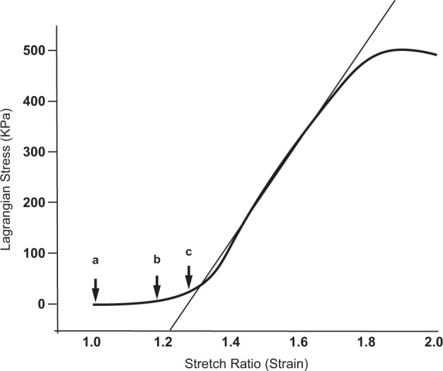
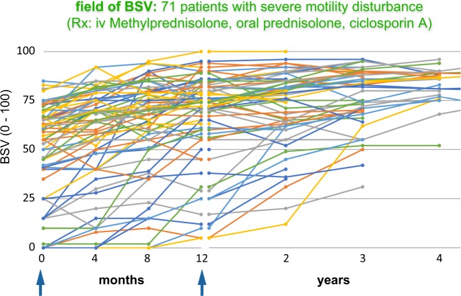
The clinical features of thyroid eye disease are dictated by the orbit's compartmentalisation; particularly, the muscle cone, which is delimited by the rectus muscles, their inter-muscular septa and the posterior sclera. The cone is anchored to the orbit apex and contains the posterior globe, the muscle bellies, a fat pad, and the blood circulation, optic nerve, and CSF sheath. It is surrounded by mobile extraconal fat, retained by the orbital septum.Thyroid eye disease is caused by expansion of muscle bellies and fat within the cone. Mechanical properties of the cone determine that the disease partitions into three phases: circumferential expansion, with forward displacement of extraconal fat; axial elongation, with increasing cone pressure; impedance of posterior venous outflow, with cone oedema and venous flow reversal.Venous flow reversal can be observed in the conjunctival circulation. It is initially transient, accompanying rises in cone pressure caused by eye movements, but later becomes permanent. It is a useful clinical sign that locates diseased muscles and anticipates venous compressive crises.Strabismus arises when inflamed rectus muscles, swollen by hydrated glycosaminoglycans, lose contractility and compliance. The incomitance is moderated by increasing stiffness affecting all the rectus muscles, as they are stretched during cone expansion.Immunomodulation, which rapidly reduces cone volume, relieving muscle elongation and stiffness, may paradoxically unmask strabismus. However, ciclosporin A suppresses late post-inflammatory fibrosis and only 4 of 71 patients so-treated required strabismus surgery.The cone model also accounts for the variety of clinical presentations of thyroid eye disease.


Conflict of interest statement

The authors declare that they have no conflict of interest.

Figures

Fig. 1
Fig. 1
The distribution of stress in the wall of a pressurised vessel depends on its shape. If it is spherical, wall stress is uniform; when it is cylindrical, circumferential stress (S2) is twice the axial stress (S1) [3]
Fig. 2
Fig. 2
Stress–strain function for normal bovine rectus muscle ex vivo (redrawn from Yoo et al. [4]). Increasing cone volume is accommodated by elongation of the rectus muscles (a, b). As the linear phase of the stress–strain curve is approached, the rectus muscles are stretched until the force they return generates cone pressure approaching that in the superior ophthalmic vein (c)
Fig. 3
Fig. 3
The cone model. Normal: the rectus muscles anchor the globe to the orbital apex. Connective tissue encapsulates the muscles and joins them (the intermuscular septa). This defines the muscle cone: formula image 
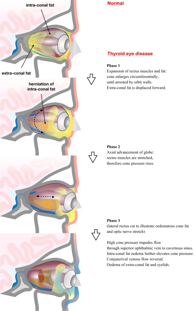
. Venous drainage to the cavernous sinus passes both through the cone (superior ophthalmic vein) and outside it (inferior ophthalmic vein). The cone is surrounded by extraconal fat. Thyroid eye disease: 1/ circumferential enlargement of the muscle cone displaces extraconal fat; 2/ stretching of rectus muscles elevates cone pressure; 3/ high pressure impedes venous drainage to the cavernous sinus.
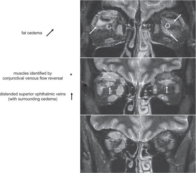
Fig. 4
Fig. 4
Venous compressive crisis: coronal STIR MRI scan (cuts immediately behind the globes, then 8 mm apart). Note the high STIR signal from swollen, inflamed rectus muscles. The muscles with overlying conjunctival venous flow reversal are shown in the middle pane (*)
Fig. 5
Fig. 5
a Unannotated. b Rectus muscles outlined (black); intermuscular septa outlined (white). Venous compressive crisis: coronal T1 MRI scan (same patient as Fig. 4: level 1 only). Note the inferotemporal herniation of cone fat: cone has expanded until its arrest by walls of the orbit
Fig. 6
Fig. 6
a Primary position. b Forced gaze away from circulation under examination stretches the underlying rectus muscle. Conjunctival veins normally flow into the limbal venous circle and episcleral collecting veins. The more stretched the muscle cone, the less compliant the muscles become. Contraction of a rectus muscle can no longer extend its antagonist sufficiently and rotation of the globe is limited. However, cone pressure rises greatly: episcleral collecting veins become compressed and conjunctival venous flow reverses
Fig. 7
Fig. 7
Conjunctival venous flow reversal over a superior rectus muscle (please see video). White arrow illustrates reversal of the normal direction of venous flow, triggered by down-gaze
Fig. 8
Fig. 8
Coronal STIR MRI during venous hypertensive crisis complicating thyroid eye disease, showing expulsion of CSF from optic nerve sheath (arrow). Patient had benign intracranial hypertension. Intraocular pressures: Right 30 / left 32mmHg. Intracranial pressure: 20 mmHg
Fig. 9
Fig. 9
Fields of BSV: 71 patients with severe motility disturbance (Rx: iv Methylprednisolone, oral prednisolone, and ciclosporin A). Incomitance arises in thyroid eye disease when particular muscles can no longer relax or contract in order to maintain BSV (due to inflammation, hydrated glycosaminoglycans, and increased muscle bulk). Stretching of the rectus muscles (which accompanies phase 2 of the disease) reduces muscle compliance globally and should, therefore, moderate such disturbances. Immunomodulation causes a rapid drop in cone pressure, negating the global component of motility restriction and lifting the suppression of myopathic strabismus. For the group as a whole, BSV began to improve immediately after immunomodulation; however, many patients deteriorated initially, as predicted. The antifibrotic effect of cyclosporin A did prevent scarring of damaged muscles during recovery, and strabismus surgery was required in only four patients

References

    1. Khong JJ, McNab AA, Ebeling PR, et al. Pathogenesis of thyroid eye disease: review and update on molecular mechanisms. Br J Ophthalmol. 2016;100:142–50. doi: 10.1136/bjophthalmol-2015-307399. - DOI - PubMed
    1. Meyer PAR. Avoiding surgery for thyroid eye disease. Eye. 2006;20:1171–7. doi: 10.1038/sj.eye.6702393. - DOI - PubMed
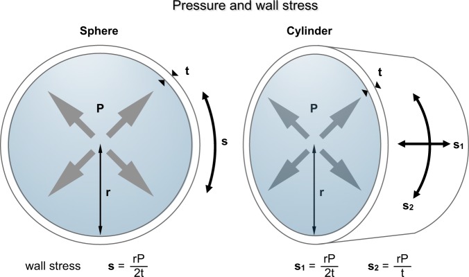
    1. Gordon JE. Structures. Penguin: UK, 1978.
    1. Yoo L, Kim H, Gupta V, Demer J. Quasilinear viscoelastic behavior of bovine extra ocular muscle tissue. Invest Ophthalmol Vis Sci. 2009;50:3721–8. doi: 10.1167/iovs.08-3245. - DOI - PMC - PubMed
    1. Frueh BR. Graves’ eye disease: orbital compliance and other physical measurements. Trans Am Ophthalmol Soc. 1984;82:492–598. - PMC - PubMed

Publication types

MeSH terms