

Physiological roles and molecular mechanisms of K+ -Cl- cotransport in the mammalian kidney and cardiovascular system: where are we?
- PMID: 30659612
- PMCID: PMC6418756
- DOI: 10.1113/JP276807
Physiological roles and molecular mechanisms of K+ -Cl- cotransport in the mammalian kidney and cardiovascular system: where are we?
Abstract
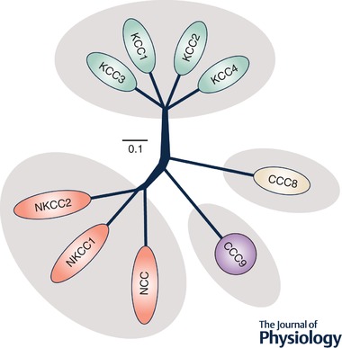
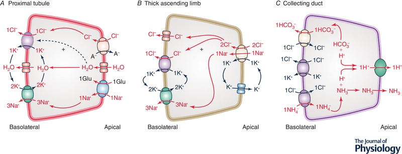
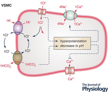
In the early 80s, renal microperfusion studies led to the identification of a basolateral K+ -Cl- cotransport mechanism in the proximal tubule, thick ascending limb of Henle and collecting duct. More than ten years later, this mechanism was found to be accounted for by three different K+ -Cl- cotransporters (KCC1, KCC3 and KCC4) that are differentially distributed along the renal epithelium. Two of these isoforms (KCC1 and KCC3) were also found to be expressed in arterial walls, the myocardium and a variety of neurons. Subsequently, valuable insights have been gained into the molecular and physiological properties of the KCCs in both the mammalian kidney and cardiovascular system. There is now robust evidence indicating that KCC4 sustains distal renal acidification and that KCC3 regulates myogenic tone in resistance vessels. However, progress in understanding the functional significance of these transporters has been slow, probably because each of the KCC isoforms is not identically distributed among species and some of them share common subcellular localizations with other KCC isoforms or sizeable conductive Cl- pathways. In addition, the mechanisms underlying the process of K+ -Cl- cotransport are still ill defined. The present review focuses on the knowledge gained regarding the roles and properties of KCCs in renal and cardiovascular tissues.
Keywords: Animal models; Cardiovascular system; Cation-Cl− cotransporter; K+-Cl− cotransporter; Renal tubular acidosis; Systemic hypertension.
© 2019 The Authors. The Journal of Physiology © 2019 The Physiological Society.
Figures
Similar articles
-
Cloning and characterization of KCC3 and KCC4, new members of the cation-chloride cotransporter gene family.J Biol Chem. 1999 Jun 4;274(23):16355-62. doi: 10.1074/jbc.274.23.16355. J Biol Chem. 1999. PMID: 10347194
-
Molecular evidence for a role for K(+)-Cl(-) cotransporters in the kidney.Am J Physiol Renal Physiol. 2013 Nov 15;305(10):F1402-11. doi: 10.1152/ajprenal.00390.2013. Epub 2013 Oct 2. Am J Physiol Renal Physiol. 2013. PMID: 24089410 Free PMC article.
-
Functional comparison of the K+-Cl- cotransporters KCC1 and KCC4.J Biol Chem. 2000 Sep 29;275(39):30326-34. doi: 10.1074/jbc.M003112200. J Biol Chem. 2000. PMID: 10913127
-
[Pathophysiological aspects of K+: Cl- cotransporters].Rev Invest Clin. 2014 Mar-Apr;66(2):173-80. Rev Invest Clin. 2014. PMID: 24960328 Review. Spanish.
-
Human and murine phenotypes associated with defects in cation-chloride cotransport.Annu Rev Physiol. 2002;64:803-43. doi: 10.1146/annurev.physiol.64.081501.155847. Annu Rev Physiol. 2002. PMID: 11826289 Review.
Cited by
-
The Integral Role of Chloride & With-No-Lysine Kinases in Cell Volume Regulation & Hypertension.Int J Nephrol Renovasc Dis. 2023 Aug 14;16:183-196. doi: 10.2147/IJNRD.S417766. eCollection 2023. Int J Nephrol Renovasc Dis. 2023. PMID: 37601040 Free PMC article. Review.
-
Molecular mechanisms, physiological roles, and therapeutic implications of ion fluxes in bone cells: Emphasis on the cation-Cl- cotransporters.J Cell Physiol. 2022 Dec;237(12):4356-4368. doi: 10.1002/jcp.30879. Epub 2022 Sep 20. J Cell Physiol. 2022. PMID: 36125923 Free PMC article. Review.
-
Molecular characteristics and physiological roles of Na+ -K+ -Cl- cotransporter 2.J Cell Physiol. 2021 Mar;236(3):1712-1729. doi: 10.1002/jcp.29997. Epub 2020 Aug 10. J Cell Physiol. 2021. PMID: 32776569 Free PMC article. Review.
-
Angiotensin II hypertension along the female rat tubule: predicted impact on coupled transport of Na+ and K.Am J Physiol Renal Physiol. 2023 Dec 1;325(6):F733-F749. doi: 10.1152/ajprenal.00232.2023. Epub 2023 Oct 12. Am J Physiol Renal Physiol. 2023. PMID: 37823196 Free PMC article.
-
Highly conserved ion binding sites are not all functionally relevant in mouse KCC4.Front Mol Biosci. 2025 Mar 31;12:1556250. doi: 10.3389/fmolb.2025.1556250. eCollection 2025. Front Mol Biosci. 2025. PMID: 40230454 Free PMC article.
References
-
- Adragna NC, Chen Y, Delpire E, Lauf PK & Morris M (2004). Hypertension in K‐Cl cotransporter‐3 knockout mice. Adv Exp Med Biol 559, 379–385. - PubMed
-
- Agalakova NI & Gusev GP (2009). Effects of phorbol 12‐myristate 13‐acetate on potassium transport in the red blood cells of frog Rana temporaria . J Comp Physiol B 179, 443–450. - PubMed
-
- Akar F, Skinner E, Klein JD, Jena M, Paul RJ & O'Neill WC (1999). Vasoconstrictors and nitrovasodilators reciprocally regulate the Na+‐K+‐2Cl− cotransporter in rat aorta. Am J Physiol 276, C1383–C1390. - PubMed
-
- Alberts B, Johnson A, Lewis J, Morgan DO, Raff MC, Roberts K, Walter P, Wilson JH & Hunt T (2015). Molecular Biology of the Cell. Garland Science, New York.
-
- Avison MJ, Gullans SR, Ogino T & Giebisch G (1988). Na+ and K+ fluxes stimulated by Na+‐coupled glucose transport: evidence for a Ba2+‐insensitive K+ efflux pathway in rabbit proximal tubules. J Membr Biol 105, 197–205. - PubMed
Publication types
MeSH terms
Substances
Grants and funding
LinkOut - more resources
Full Text Sources