. 2019 Mar;17(3):482-491.
doi: 10.1111/jth.14389. Epub 2019 Feb 20.

Virus envelope tissue factor promotes infection in mice

Affiliations

Virus envelope tissue factor promotes infection in mice

Michael R Sutherland et al. J Thromb Haemost. 2019 Mar.

Abstract

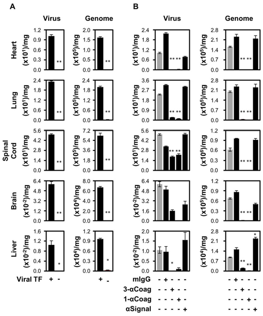
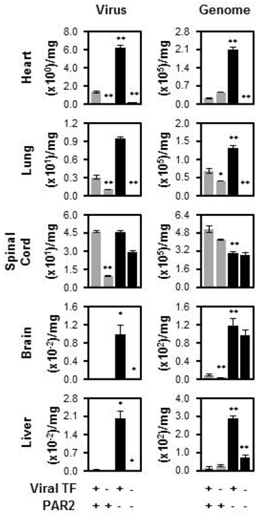
Essentials The coagulation initiator, tissue factor (TF), is on the herpes simplex virus 1 (HSV1) surface. HSV1 surface TF was examined in mice as an antiviral target since it enhances infection in vitro. HSV1 surface TF facilitated infection of all organs evaluated and anticoagulants were antiviral. Protease activated receptor 2 inhibited infection in vivo and its pre-activation was antiviral. SUMMARY: Background Tissue factor (TF) is the essential cell surface initiator of coagulation, and mediates cell signaling through protease-activated receptor (PAR) 2. Having a diverse cellular distribution, TF is involved in many biological pathways and pathologies. Our earlier work identified host cell-derived TF on the envelope covering several viruses, and showed its involvement in enhanced cell infection in vitro. Objective In the current study, we evaluated the in vivo effects of virus surface TF on infection and on the related modulator of infection PAR2. Methods With the use of herpes simplex virus type 1 (HSV1) as a model enveloped virus, purified HSV1 was generated with or without envelope TF through propagation in a TF-inducible cell line. Infection was studied after intravenous inoculation of BALB/c, C57BL/6J or C57BL/6J PAR2 knockout mice with 5 × 10⁵ plaque-forming units of HSV1, mimicking viremia. Three days after inoculation, organs were processed, and virus was quantified with plaque-forming assays and quantitative real-time PCR. Results Infection of brain, lung, heart, spinal cord and liver by HSV1 required viral TF. Demonstrating promise as a therapeutic target, virus-specific anti-TF mAbs or small-molecule inhibitors of coagulation inhibited infection. PAR2 modulates HSV1 in vivo as demonstrated with PAR2 knockout mice and PAR2 agonist peptide. Conclusion TF is a constituent of many permissive host cell types. Therefore, the results presented here may explain why many viruses are correlated with hemostatic abnormalities, and indicate that TF is a novel pan-specific envelope antiviral target.

Keywords: anticoagulant; coagulation; protease-activated receptor; tissue factor; virus infection.


Conflict of interest statement

Disclosure of Conflicts of Interests

The authors state that they have no conflict of interest.

Figures

Fig. 1.
Fig. 1.
Infection of Th2 immuno-dominant mice (BALB/c) is enhanced by TF on HSV1. (A) Eight-week-old female BALB/c mice were inoculated intravenously with 5 × 10⁵ PFU of either TF competent (TF+; n=24) or deficient (TF−; n=13) HSV1 via the tail vein. Three days post-infection, the mice were processed and the amount of infectious virus (Virus; PFU/mg) and HSV1 genetic material (Genome; genome copies/mg) were determined. (B) Additional experiments were conducted after pre-immunization of mice with a mixture of three anti-TF IgG1 monoclonal antibodies, 5G9, 9C3 and 6B4 (3-αCoag; at 0.33 mg each/mouse; n=10), 5G9 alone (1-αCoag; 1 mg/mouse; n=6) or 10H10 alone (αSignal; 1 mg/mouse; n=6), 4 hours prior to injection of the virus. Purified anti-Simian virus 40 large T antigen (mIgG; 1 mg/mouse; n=8) was used as an irrelevant monoclonal IgG1 subclass-matched negative control. Grey bars represent data from HSV1/TF+ as depicted in Panel A. In all panels data are expressed as mean ± SEM. As determined by Student’s t test, **P ≤ 0.05 and *P ≤ 0.10 when compared to the TF+ virus.
Fig. 2.
Fig. 2.
Infection of BALB/c mice is inhibited by anticoagulation. Eight-week-old female BALB/c mice were inoculated intravenously with 5 × 10⁵ PFU of TF competent (n=24) HSV1 alone (grey bar as depicted in Figure 1, Panel A) or simultaneously with hirudin (1 mg/kg, n=9), NAPc2 (1 mg/kg, n=18), or apixaban (1 mg/kg, n=18) via the tail vein. In each case, three days post infection, the mice were processed and the amount of infectious virus (PFU/mg) was determined in each organ. In all panels data are expressed as mean ± SEM. As determined by Student’s t test, **P ≤ 0.05 and *P ≤ 0.10 when compared to the TF+ virus.
Fig. 3.
Fig. 3.
Infection of Th1 immuno-dominant mice (C57BL/6J) is enhanced by TF on HSV1. Eight week old female C57BL/6J mice were inoculated intravenously with 5 × 10⁵ PFU of either TF competent (TF+, n=7) or deficient (TF−; n=8) HSV1 via the tail vein. Three days post-infection, the mice were processed and the amount of infectious virus (Virus; PFU/mg) and HSV1 genetic material (Genome; genome copies/mg) were determined. In all panels data are expressed as mean ± SEM. As determined by Student’s t test, **P ≤ 0.05 and *P ≤ 0.10 when compared to the TF+ virus.
Fig. 4.
Fig. 4.
Infection of C57BL/6J is enhanced by PAR2 deficiency. Wild type C57BL/6J mice inoculated with either TF competent (TF+, n=7, grey bar, as in Fig. 3, TF+) or deficient (TF−; n=8, grey bar, as in Fig. 3, TF−) HSV1 via the tail vein were compared to PAR2 KO (PAR2−; n=6) mice and the combined effect of PAR2 knock out and viral TF-deficiency (n=5). Three days post-inoculation, the mice were processed and the amount of infectious virus (Virus; PFU/mg) or genome copies (Genome; genome copies/mg) was determined. In all panels data are expressed as mean ± SEM. As determined by Student’s t test, **P ≤ 0.05 and *P ≤ 0.10 and when compared to the TF+ virus except for the TF−/PAR2− which is compared to the TF− virus.
Fig. 5.
Fig. 5.
Infection of BALB/c mice is inhibited by PAR2 activating peptide. Experiments were conducted as in Fig. 1 using BALB/c mice to investigate the simultaneous injection of TF competent HSV1 (grey bar, from Fig. 1A, Virus, TF+) with PAR2ap (3 umol/kg, n=7). In all panels data are expressed as mean ± SEM. As determined by Student’s t test, **P ≤ 0.05 and *P ≤ 0.10 compared to no addition of PAR2ap.

References

    1. Rosen ED, Chan JC, Idusogie E, Clotman F, Vlasuk G, Luther T, Jalbert LR, Albrecht S, Zhong L, Lissens A, Schoonjans L, Moons L, Collen D, Castellino FJ, Carmeliet P. Mice lacking factor VII develop normally but suffer fatal perinatal bleeding. Nature 1997;390:290–294. - PubMed
    1. Carmeliet P, Mackman N, Moons L, Luther T, Gressens P, Van V I, Demunck H, Kasper M, Breier G, Evrard P, Muller M, Risau W, Edgington T, Collen D Role of tissue factor in embryonic blood vessel development. Nature 1996;383:73–75. - PubMed
    1. Butenas S Tissue factor structure and function. Scientifica (Cairo) 2012;2012:964862. - PMC - PubMed
    1. Xu Z, Xu H, Ploplis VA, Castellino FJ. Factor VII deficiency impairs cutaneous wound healing in mice. Mol Med 2010;16:167–176. - PMC - PubMed
    1. Eisenreich A, Zakrzewicz A, Huber K, Thierbach H, Pepke W, Goldin-Lang P, Schultheiss HP, Pries A, Rauch U. Regulation of pro-angiogenic tissue factor expression in hypoxia-induced human lung cancer cells. Oncol Rep 2013;30:462–470. - PubMed

Publication types

MeSH terms