. 2019 Apr 1:189:267-275.
doi: 10.1016/j.neuroimage.2019.01.037. Epub 2019 Jan 16.

Modulation of brain function by targeted delivery of GABA through the disrupted blood-brain barrier

Affiliations

Modulation of brain function by targeted delivery of GABA through the disrupted blood-brain barrier

Nick Todd et al. Neuroimage. .

Abstract

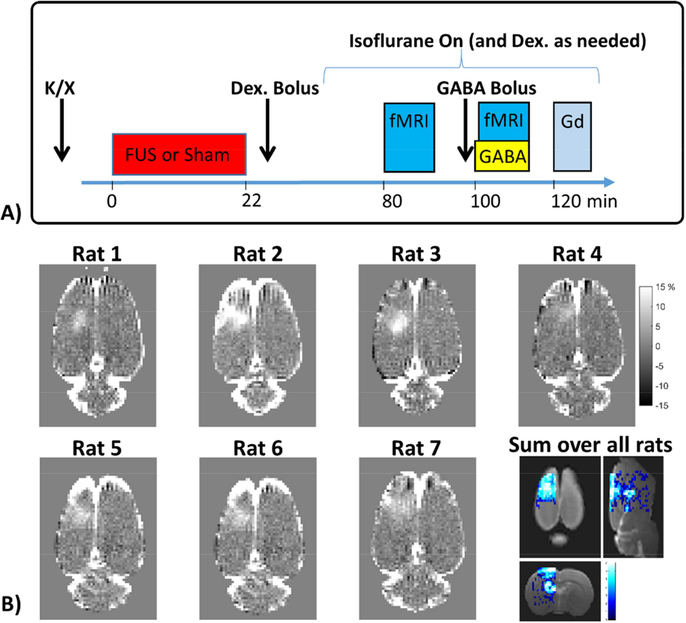
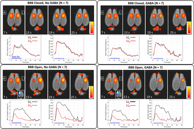
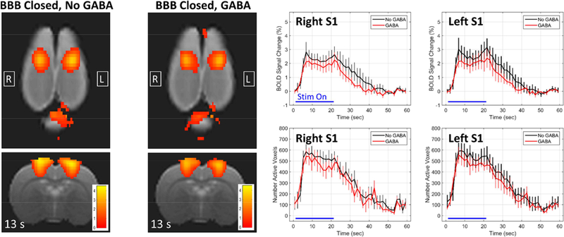
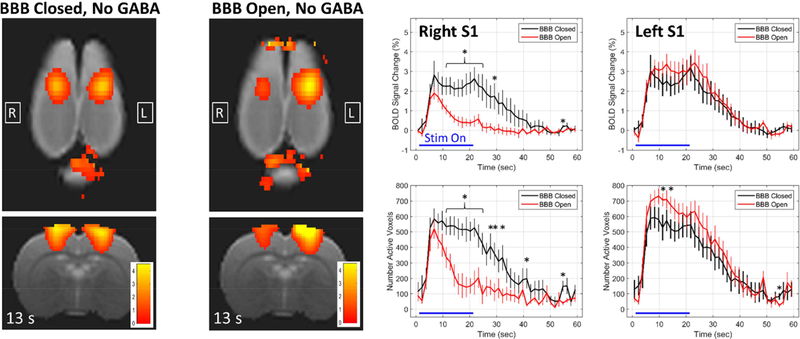
The technology of transcranial focused ultrasound (FUS) enables a novel approach to neuromodulation, a tool for selective manipulation of brain function to be used in neurobiology research and with potential applications in clinical treatment. The method uses transcranial focused ultrasound to non-invasively open the blood-brain barrier (BBB) in a localized region such that a systemically injected neurotransmitter chemical can be delivered to the targeted brain site. The approach modulates the chemical signaling that occurs in and between neurons, making it complimentary to most other neuromodulation techniques that affect the electrical properties of neuronal activity. Here, we report delivering the inhibitory neurotransmitter GABA to the right somatosensory cortex of the rat brain during bilateral hind paw electrical stimulation and measure the inhibition of activation using functional MRI (fMRI). In a 2 × 2 factorial design, we evaluated conditions of BBB Closed vs BBB Open and No GABA vs GABA. Results from fMRI measurements of the blood oxygen level-dependent (BOLD) signal show: 1) intravenous GABA injection without FUS-mediated BBB opening does not have an effect on the BOLD response; 2) FUS-mediated BBB opening alone significantly alters the BOLD signal response to the stimulus, both in amplitude and shape of the time course; 3) the combination of FUS-mediated BBB opening and GABA injection further reduces the peak amplitude and spatial extent of the BOLD signal response to the stimulus. The data support the thesis that FUS-mediated opening of the BBB can be used to achieve non-invasive delivery of neuroactive substances for targeted manipulation of brain function.

Keywords: Blood-brain barrier; Brain; Brain networks; Drug delivery; Focused ultrasound; Functional MRI; Neuromodulation.


Conflict of interest statement

Competing Interests

The authors declare that they have no financial or non-financial competing interests related to this work.

Figures

Figure 1:
Figure 1:. Experiment timing and BBB opening.
A) Timing of an experimental session, which consists of ketamine/xylazine injection, FUS sonications (for BBB Open cases) or Sham FUS (for BBB Closed cases), injection of the Dexdomitor bolus and a waiting period to transfer the rat to the fMRI imaging set up and allow for physiological stabilization, the fMRI runs without and with GABA injection, and finally gadolinium contrast imaging to assess BBB opening. B) T1-weighted contrast images (converted to percent difference) showing the BBB opening for each individual rat and a map of the sum of binary opening images over all rats overlaid on a T2w anatomical image.
Figure 2:
Figure 2:. Summary of results over the four experimental conditions.
Maps of BOLD signal percent change are shown at four different time points for each of the four experimental conditions. Corresponding plots of BOLD signal change and number of active voxels are shown for the left and right S1 regions (mean over all rats).
Figure 3:
Figure 3:. Effects of GABA when the BBB is closed.
Maps of BOLD signal change at 13 seconds into the stimulation block are shown for the two conditions of BBB Closed, No GABA and BBB Closed, GABA. The plots compare the BBB Closed, No GABA condition (black) against the BBB Closed, GABA condition (red) for the right and left S1 regions (mean +/− standard error). No significant differences exist at any time point for either the BOLD change or the number of active voxels in either the right or left S1.
Figure 4:
Figure 4:. Changes in the BOLD signal response due to BBB opening alone.
Maps of BOLD signal change at 13 seconds into the stimulation block are shown for the two conditions of BBB Closed, No GABA and BBB Open, No GABA. The plots compare the BBB Closed, No GABA condition (black) against the BBB Open, No GABA condition (red) for the right and left S1 regions (mean +/− standard error). * indicates time points at which the two conditions are significantly different (p < 0.05).
Figure 5:
Figure 5:. Histology after BBB opening.
The two row shows the T1w contrast difference image and hematoxylin and eosin staining from two different rats. Regions from the right and left somatosensory cortex are shown at 1.25x, 10x, and 40x magnification. The 10x and 40x images of the right cortex where taken from a region that displayed hyperintensity on the T1w contrast difference images. No signs of damage to neuronal cells were seen in either rat.
Figure 6:
Figure 6:. Effects of BBB opening combined with GABA delivery.
Maps of BOLD signal change at 7 seconds into the stimulation block are shown for the two conditions of BBB Open, No GABA and BBB Open, GABA. The plots compare the BBB Open, No GABA condition (black) against the BBB Open, GABA condition (red) for the right and left S1 regions (mean +/− standard error). * indicates time points at which the two conditions are significantly different (p < 0.05).
Figure 7.
Figure 7.
Correlation of GABA effects with extent of BBB opening. The difference in BOLD change values between the left S1 and right S1 regions were calculated for each individual rat. These values were correlated with the extent of BBB opening seen in each individual rat, as measured by the mean gadolinium percent change in the right S1 ROI. The first two panels show scatter plots of the data at two different time points (t = 7 seconds and t = 16 seconds). The third panel shows a plot of correlation values for each time point in the stimulation block. Strong positive correlation indicates that lower activation seen in the right S1 compared to the left S1 was associated with stronger BBB opening.

References

    1. Adamczak JM, Farr TD, Seehafer JU, Kalthoff D, Hoehn M, 2010. High field BOLD response to forepaw stimulation in the mouse. Neuroimage 51, 704–712. 10.1016/j.neuroimage.2010.02.083 - DOI - PubMed
    1. Alborch E, Torregrosa G, Terrasa JC, Estrada C, 1984. GABA receptors mediate cerebral vasodilation in the unanesthetized goat. Brain Res. 321, 103–110. 10.1016/0006-8993(84)90685-1 - DOI - PubMed
    1. Armbruster BN, Li X, Pausch MH, Herlitze S, Roth BL, 2007. Evolving the lock to fit the key to create a family of G protein-coupled receptors potently activated by an inert ligand. Proc. Natl. Acad. Sci 104, 5163–5168. 10.1073/pnas.0700293104 - DOI - PMC - PubMed
    1. Aryal M, Arvanitis CD, Alexander PM, McDannold N, 2014. Ultrasound-mediated blood-brain barrier disruption for targeted drug delivery in the central nervous system. Adv. Drug Deliv. Rev 72, 94–109. 10.1016/j.addr.2014.01.008 - DOI - PMC - PubMed
    1. Boyden ES, Zhang F, Bamberg E, Nagel G, Deisseroth K, 2005. Millisecond-timescale, genetically targeted optical control of neural activity. Nat. Neurosci 8, 1263–8. 10.1038/nn1525 - DOI - PubMed

Publication types

Substances