

Creation of a Porcine Kyphotic Model
- PMID: 30660214
- PMCID: PMC7341554
- DOI: 10.1016/j.jspd.2018.07.002
Creation of a Porcine Kyphotic Model
Abstract
Study design: Large animal study.
Objective: Create a thoracic hyperkyphotic deformity in an immature porcine spine, so that future researchers may use this model to validate spinal instrumentation and other therapies used in the treatment of hyperkyphosis.
Summary of background data: Although several scoliotic animal models have been developed, there have been no reports of a thoracic hyperkyphotic animal model creation in an immature animal. The present study was designed to produce a porcine hyperkyphotic model by the time the pig weighed 25 kg, which corresponds to the approximate weight of a child undergoing surgery for early-onset scoliosis (EOS).
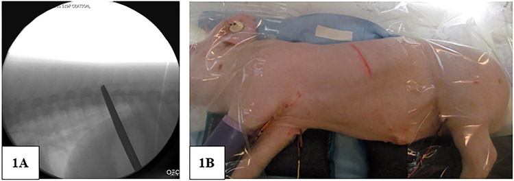
Methods: Successful surgical procedures were performed in 6 consecutive 10-kg (male, 5-week-old) immature Yorkshire pigs. Procedure protocol consisted of 1) a left thoracotomy at T10-T11, 2) screw placement at T9 and T11, 3) partial vertebrectomy at T10, 4) posterior interspinous ligament transection, and 5) placement of wire loop around screws and tightening. Weekly x-ray imaging was performed preoperatively and postoperatively, documenting progressively increasing kyphosis as the pig grew. Necropsy was performed 5-6 weeks after surgery, with CT, slab section, and histologic analysis.
Results: Average T9-T11 kyphosis (measured by sagittal Cobb angle) was 6.1° ± 1.4° (mean ± SD) preoperatively, 30.5° ± 1.0° immediately postoperation, and significantly increased to 50.3° ± 7.2° (p < .0001) over 5-6 weeks in 6 consecutive pigs at time of necropsy.
Conclusions: An animal model of relatively more rigid-appearing thoracic hyperkyphotic deformities in immature pigs has been created. Subsequent studies addressing management of early-onset kyphosis with spinal instrumentation are now possible.
Level of evidence: Level V.
Keywords: Animal model; Early-onset scoliosis; Immature spine; Pig; Thoracic hyperkyphosis.
Copyright © 2018 Scoliosis Research Society. Published by Elsevier Inc. All rights reserved.
Conflict of interest statement
Conflict of Interest:
None of the authors of this paper have a conflict of interest that might be construed as affecting the conduct or reporting of the work presented.
Figures


References
-
- Lonner B, Toombs CS, Guss M, et al. Complications in operative Scheuermann Kyphosis. Spine 2015;40:305–11. - PubMed
-
- Schroerlucke SR, Akbarnia BA, Pawelek JB, et al. How does thoracic kyphosis affect patient outcomes in growing rod surgery? Spine 2012;37:1303–9. - PubMed
-
- Watanabe K, Uno K, Suzuki T, et al. Risk factors for complications associated with growing-rod surgery for early-onset scoliosis. Spine 2013;38:E464–8. - PubMed
-
- Langenskiold A, Michelsson JE. The pathogenesis of experimental progressive scoliosis. Acta orthopaedica Scandinavica. Supplementum 1962;59:1–26. - PubMed
-
- Bobyn JD, Little DG, Gray R, et al. Animal models of scoliosis. J Orthop Res 2015;33:458–67. - PubMed
Publication types
MeSH terms
Grants and funding
LinkOut - more resources
Full Text Sources
Research Materials