

Should We Resect and Discard Low Risk Diminutive Colon Polyps
- PMID: 30661337
- PMCID: PMC6547333
- DOI: 10.5946/ce.2018.136
Should We Resect and Discard Low Risk Diminutive Colon Polyps
Abstract
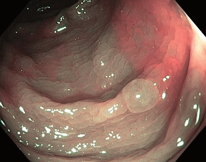
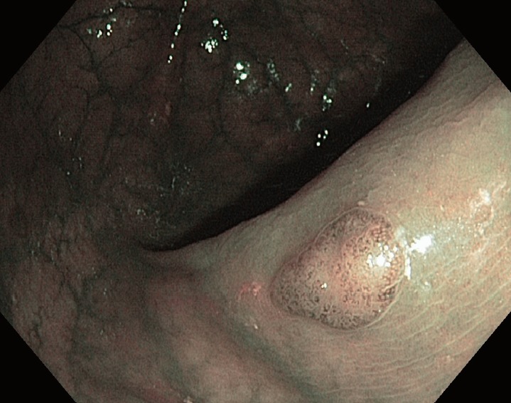
Diminutive colorectal polyps <5 mm are very common and almost universally benign. The current strategy of resection with histological confirmation of all colorectal polyps is costly and may increase the risk of colonoscopy. Accurate, optical diagnosis without histology can be achieved with currently available endoscopic technologies. The American Society of Gastrointestinal Endoscopy Preservation and Incorporation of Valuable endoscopic Innovations supports strategies for optical diagnosis of small non neoplastic polyps as long as two criteria are met. For hyperplastic appearing polyps <5 mm in recto-sigmoid colon, the negative predictive value should be at least 90%. For diminutive low grade adenomatous appearing polyps, a resect and discard strategy should be sufficiently accurate such that post-polypectomy surveillance recommendations based on the optical diagnosis, agree with a histologically diagnosis at least 90% of the time. Although the resect and discard as well as diagnose and leave behind approach has major benefits with regard to both safety and cost, it has yet to be used widely in practice. To fully implement such as strategy, there is a need for better-quality training, quality assurance, and patient acceptance. In the article, we will review the current state of the science on optical diagnose of colorectal polyps and its implications for colonoscopy practice.
Keywords: Colorectal polyps.
Conflict of interest statement
Figures
Similar articles
-
Simplifying Resect and Discard Strategies for Real-Time Assessment of Diminutive Colorectal Polyps.Clin Gastroenterol Hepatol. 2018 May;16(5):706-714. doi: 10.1016/j.cgh.2017.11.036. Epub 2017 Nov 23. Clin Gastroenterol Hepatol. 2018. PMID: 29174789
-
Advances in the removal of diminutive colorectal polyps.Expert Rev Gastroenterol Hepatol. 2015 Feb;9(2):237-44. doi: 10.1586/17474124.2014.950955. Epub 2014 Aug 26. Expert Rev Gastroenterol Hepatol. 2015. PMID: 25155348 Review.
-
Optical diagnosis of diminutive polyps in the Dutch Bowel Cancer Screening Program: Are we ready to start?Endosc Int Open. 2020 Mar;8(3):E257-E265. doi: 10.1055/a-1072-4853. Epub 2020 Feb 21. Endosc Int Open. 2020. PMID: 32118099 Free PMC article.
-
New paradigms for colonoscopic management of diminutive colorectal polyps: predict, resect, and discard or do not resect?Clin Endosc. 2013 Mar;46(2):130-7. doi: 10.5946/ce.2013.46.2.130. Epub 2013 Mar 31. Clin Endosc. 2013. PMID: 23614122 Free PMC article.
-
Optical Technologies for Endoscopic Real-Time Histologic Assessment of Colorectal Polyps: A Meta-Analysis.Am J Gastroenterol. 2019 Aug;114(8):1219-1230. doi: 10.14309/ajg.0000000000000156. Am J Gastroenterol. 2019. PMID: 30848728 Review.
Cited by
-
Colon capsule endoscopy is an effective filter test for colonic polyp surveillance.World J Gastrointest Endosc. 2025 May 16;17(5):101322. doi: 10.4253/wjge.v17.i5.101322. World J Gastrointest Endosc. 2025. PMID: 40438717 Free PMC article.
-
UEG Week 2019 Poster Presentations.United European Gastroenterol J. 2019 Oct;7(8_suppl):189-1030. doi: 10.1177/2050640619854671. United European Gastroenterol J. 2019. PMID: 32213001 Free PMC article. No abstract available.
-
Too Good to Be True? Evaluation of Colonoscopy Sensitivity Assumptions Used in Policy Models.Cancer Epidemiol Biomarkers Prev. 2022 Apr 1;31(4):775-782. doi: 10.1158/1055-9965.EPI-21-1001. Cancer Epidemiol Biomarkers Prev. 2022. PMID: 34906968 Free PMC article.
-
Editors' Choice of Noteworthy Clinical Endoscopy Publications in the First Decade.Clin Endosc. 2021 Sep;54(5):633-640. doi: 10.5946/ce.2021.216. Epub 2021 Sep 13. Clin Endosc. 2021. PMID: 34510862 Free PMC article.
-
Non-optical polyp-based resect and discard strategy: A prospective clinical study.World J Gastroenterol. 2022 May 21;28(19):2137-2147. doi: 10.3748/wjg.v28.i19.2137. World J Gastroenterol. 2022. PMID: 35664039 Free PMC article.
References
-
- Rex DK, Overhiser AJ, Chen SC, Cummings OW, Ulbright TM. Estimation of impact of American College of Radiology recommendations on CT colonography reporting for resection of high-risk adenoma findings. Am J Gastroenterol. 2009;104:149–153. - PubMed
-
- Butterly LF, Chase MP, Pohl H, Fiarman GS. Prevalence of clinically important histology in small adenomas. Clin Gastroenterol Hepatol. 2006;4:343–348. - PubMed
Publication types
LinkOut - more resources
Full Text Sources
Medical
Miscellaneous
