. 2019 Feb 7;104(2):246-259.
doi: 10.1016/j.ajhg.2018.12.014. Epub 2019 Jan 17.

De Novo SOX4 Variants Cause a Neurodevelopmental Disease Associated with Mild Dysmorphism

Affiliations

De Novo SOX4 Variants Cause a Neurodevelopmental Disease Associated with Mild Dysmorphism

Ash Zawerton et al. Am J Hum Genet. .

Erratum in

  • De Novo SOX4 Variants Cause a Neurodevelopmental Disease Associated with Mild Dysmorphism.
    Zawerton A, Yao B, Yeager JP, Pippucci T, Haseeb A, Smith JD, Wischmann L, Kühl SJ, Dean JCS, Pilz DT, Holder SE; Deciphering Developmental Disorders Study; University of Washington Center for Mendelian Genomics; McNeill A, Graziano C, Lefebvre V. Zawerton A, et al. Am J Hum Genet. 2019 Apr 4;104(4):777. doi: 10.1016/j.ajhg.2019.01.014. Am J Hum Genet. 2019. PMID: 30951678 Free PMC article. No abstract available.

Abstract

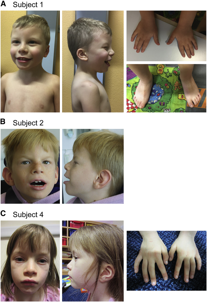
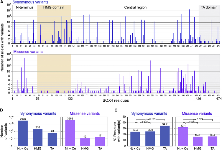
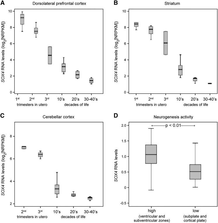
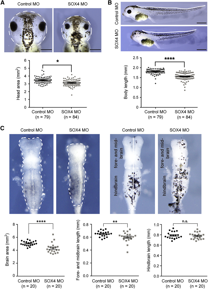
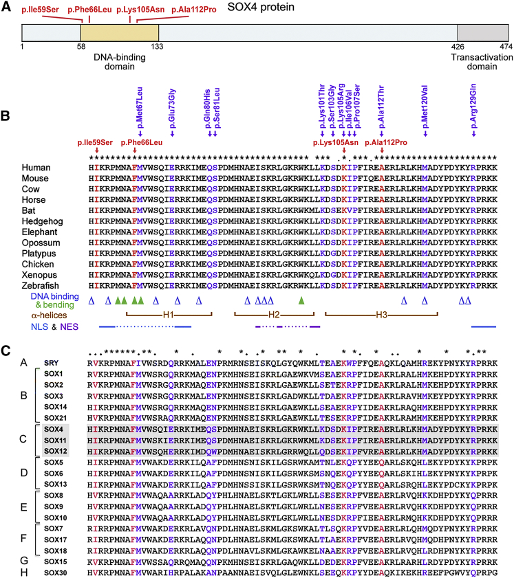
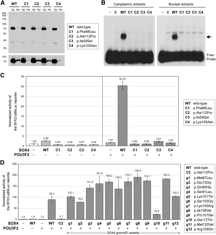
SOX4, together with SOX11 and SOX12, forms group C of SRY-related (SOX) transcription factors. They play key roles, often in redundancy, in multiple developmental pathways, including neurogenesis and skeletogenesis. De novo SOX11 heterozygous mutations have been shown to cause intellectual disability, growth deficiency, and dysmorphic features compatible with mild Coffin-Siris syndrome. Using trio-based exome sequencing, we here identify de novo SOX4 heterozygous missense variants in four children who share developmental delay, intellectual disability, and mild facial and digital morphological abnormalities. SOX4 is highly expressed in areas of active neurogenesis in human fetuses, and sox4 knockdown in Xenopus embryos diminishes brain and whole-body size. The SOX4 variants cluster in the highly conserved, SOX family-specific HMG domain, but each alters a different residue. In silico tools predict that each variant affects a distinct structural feature of this DNA-binding domain, and functional assays demonstrate that these SOX4 proteins carrying these variants are unable to bind DNA in vitro and transactivate SOX reporter genes in cultured cells. These variants are not found in the gnomAD database of individuals with presumably normal development, but 12 other SOX4 HMG-domain missense variants are recorded and all demonstrate partial to full activity in the reporter assay. Taken together, these findings point to specific SOX4 HMG-domain missense variants as the cause of a characteristic human neurodevelopmental disorder associated with mild facial and digital dysmorphism.

Keywords: QA keywords.


Figures

Figure1
Figure1
Pictures of Three Subjects Subject 1 at 4 years and 8 months (A), subject 2 at 6 years and 8 months (B), and subject 4 at 6 years and 10 months (C). Frontal and profile views of the heads show mild facial dysmorphism,including anteverted nares, wide mouth with acupid bow, and posteriorly rotated ears. Pictures of the hands of subjects 1 and 4 show bilateral 5th finger clinodactyly. A picture of the feet of subject 1 shows normal morphology.
Figure 2
Figure 2
Analysis of SOX4 Missense Variants in the gnomAD Control Cohort (A) Bar graphs showing the numbers of synonymous and missense SOX4 variants detected in gnomAD for each amino acid of the SOX4 protein sequence. The HMG domain is highlighted in yellow and the transactivation domain in gray. (B) Comparison of the numbers of synonymous and missense gnomAD variants in the different SOX4 domains. Columns correspond to the N-terminal and central region (Nt + Ce), HMG domain (HMG), and transactivation domain (TA). Values are indicated at the top of each column. (C) Percentages of residues per SOX region that feature at least one gnomAD variant. The p values obtained in two-sample t tests for the comparison of regions is shown.
Figure 3
Figure 3
SOX4 Transcript Levels in the Developing and Adult Human Brain (A–C) Changes in SOX4 expression levels during development and adult life in the dorsolateral prefrontal cortex, striatum, and cerebellar cortex, as assessed by RNA-seq. The first three samples were obtained during the three trimesters of fetal development, and the next three during the first four decades of life, as indicated on the x axis. No data were available for the cerebellar cortex in the first trimester of embryogenesis. (D) RNA microarray data demonstrating that SOX4 expression is significantly higher in neuroanatomical regions with high-level neurogenesis (ventricular and subventricular zones) than in regions with low-level neurogenesis (subplate and cortical plate) at 21 weeks of gestation (Mann-Whitney U-test, p < 0.01). Error bars in the standard box plots from PASW/SPSS represent the interquartile range.
Figure 4
Figure 4
Effect of sox4 Knockdown on Xenopus laevis Embryo Development (A) Top, representative pictures of stage-43 Xenopus embryos showing that bilateral injection of sox4 MO leads to a smaller head area (white dotted circles) and to microphthalmia compared to bilateral injection of control MO. Bottom, graph showing quantification of the head area for all tested embryos. n, number of embryos. The p value was calculated by a non-parametric Mann-Whitney rank sum test. p ≤ 0.05. Scale bar, 1 mm. (B) Sox4 depletion results in shorter body length. Data are presented as in (A). ∗∗∗∗p ≤ 0.0001. Scale bar, 1 mm. (C) Sox4 deficiency impairs brain development. Top left, pictures showing that sox4 MO injections lead to a small brain area (dotted line). Top right, pictures showing that Sox4 depletion results in underdevelopment of the fore- and mid-brain, but not hindbrain (white vertical lines separated by a horizontal dotted line). Bottom panels, data quantification performed as in (A). n, number of embryos. ∗∗∗∗p ≤ 0.0001; ∗∗p ≤ 0.01; n.s., not significant.
Figure 5
Figure 5
Analysis of the Location of SOX4 Missense Variants Detected in Subjects and in gnomAD Individuals (A) Schematic of the human SOX4 protein showing the location of the four subject missense variants in the HMG domain. Variants are shown in red and their positions are marked with bars. Numbers indicate the position of amino acids in the protein sequence. (B) Alignment of the SOX4 HMG-domain sequences from various vertebrate species. Sequence accession numbers are listed in Table S1. SOX4 variants found in the study subjects and in the gnomAD control cohort are shown in red and purple, respectively, above the sequences, and amino acids matching the variants are similarly colored in the aligned sequences. Above the sequences, symbols denote fully conserved (asterisks) and semi-conserved (dots) amino acids. Below the sequences, residues important for DNA binding and bending are shown with blue open triangles and green closed triangles, respectively. Brown brackets demarcate the three α helices (H1, H2, and H3) that secondarily structure the DNA-binding domain. Key amino acids in the N-terminal and C-terminal nuclear localization signal sequences (NLS) and nuclear export signal sequence (NES) are shown with continued lines and are linked with dotted lines. (C) Comparison of the HMG domain sequences of all human SOX proteins. Sequence accession numbers are listed in Table S2.
Figure 6
Figure 6
Consequences of Missense Mutations on SOX4 Protein Level and Activity (A) Western blot of cytoplasmic (cy) and nuclear (nu) extracts from COS-1 cells transiently transfected with expression plasmids encoding wild-type (WT) or variant SOX4 proteins (C1 to C4) fused at the N terminus with a 3FLAG epitope. The proteins were identified using anti-FLAG antibody. The Mr of protein standards is indicated in k units. Note that each SOX4 protein exhibited the expected Mr of approximately 75 k. The bottom panel is a longer exposure of the same blot as in the top panel. It is limited to the SOX4 protein region and shows that SOX4 protein was present in all nuclear extracts, but at a lower level than in cytoplasmic extracts. Variant SOX4 proteins did not show significant differences in nuclear localization compared to wild-type SOX4 when several independent experiments were considered. (B) EMSA comparing the binding efficiency of SOX4 wild-type and variants. The cell extracts were the same as in (A), but also included negative controls without SOX4 (−, no plasmid; E, empty plasmid). The arrow indicates the complex formed between wild-type SOX4 and the DNA probe. (C) Assay of the ability of the four case subjects’ SOX4 variants to activate transcription. COS-1 cells were transiently transfected with a 6FXO-p89-Luc reporter, a pSV-beta-galactosidase plasmid, and expression plasmids for wild-type (WT) or variant SOX4 (C1 to C4), and POU3F2. Reporter activities are presented as the mean ± standard deviation obtained from triplicates for each condition. They were normalized for transfection efficiency and are reported as fold increase relative to the activity of the reporter in the absence of SOX4 and POU3F2. The presence (+) or absence (−) of SOX4 and POU3F2 plasmid is indicated beneath the bars. These data were reproduced in more than three independent experiments. (D) Assay of the ability of the 12 gnomAD SOX4 HMG-domain missense variants (g1 to g12) to activate transcription. COS-1 cells were transfected as described in (C) and using SOX4 and POU3F2 expression plasmids as shown in the figure. Data were calculated and are presented as in (C). They were reproduced in four independent experiments. A western blot showing that similar amounts of SOX4 protein were made in all conditions is presented in Figure S3.

References

    1. Urbán N., Guillemot F. Neurogenesis in the embryonic and adult brain: same regulators, different roles. Front. Cell. Neurosci. 2014;8:396. - PMC - PubMed
    1. Chen C., Lee G.A., Pourmorady A., Sock E., Donoghue M.J. Orchestration of neuronal differentiation and progenitor pool expansion in the developing cortex by SoxC genes. J. Neurosci. 2015;35:10629–10642. - PMC - PubMed
    1. Lefebvre V., Bhattaram P. Vertebrate skeletogenesis. Curr. Top. Dev. Biol. 2010;90:291–317. - PMC - PubMed
    1. Kozhemyakina E., Lassar A.B., Zelzer E. A pathway to bone: signaling molecules and transcription factors involved in chondrocyte development and maturation. Development. 2015;142:817–831. - PMC - PubMed
    1. Warman M.L., Cormier-Daire V., Hall C., Krakow D., Lachman R., LeMerrer M., Mortier G., Mundlos S., Nishimura G., Rimoin D.L. Nosology and classification of genetic skeletal disorders: 2010 revision. Am. J. Med. Genet. A. 2011;155A:943–968. - PMC - PubMed

Publication types

MeSH terms