

Glycerol kinase 2 is essential for proper arrangement of crescent-like mitochondria to form the mitochondrial sheath during mouse spermatogenesis
- PMID: 30662012
- PMCID: PMC6473107
- DOI: 10.1262/jrd.2018-136
Glycerol kinase 2 is essential for proper arrangement of crescent-like mitochondria to form the mitochondrial sheath during mouse spermatogenesis
Abstract
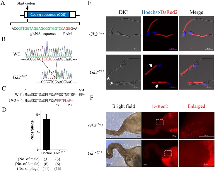
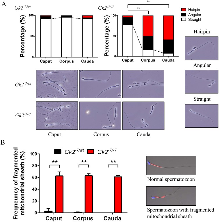
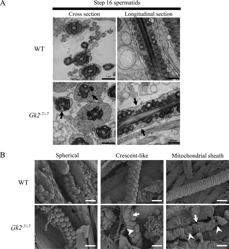
The mitochondrial sheath is composed of mitochondria that coil tightly around the midpiece of sperm flagellum. These mitochondria are recruited from the cytoplasm to the flagellum late in spermatogenesis. Initially, recruited mitochondria are spherical-shaped but then elongate laterally to become crescent-like in shape. Subsequently, crescent-like mitochondria elongate continuously to coil tightly around the flagellum. Recently, disorganization of the mitochondrial sheath was reported in Glycerol kinase 2 (Gk2) disrupted mice. To analyze the disorganization of the mitochondrial sheath further, we generated Gk2-deficient mice using the CRISPR/Cas9 system and observed sperm mitochondria in testis using a freeze-fracture method with scanning electron microscopy. Gk2-disrupted spermatids show abnormal localization of crescent-like mitochondria, in spite of the initial proper alignment of spherical mitochondria around the flagellum, which causes abnormal mitochondrial sheath formation leading to exposure of the outer dense fibers. These results indicate that GK2 is essential for proper arrangement of crescent-like mitochondria to form the mitochondrial sheath during mouse spermatogenesis.
Keywords: Glycerol kinase; Male infertility; Mitochondrial sheath formation; Sperm mitochondria; Spermatogenesis.
Figures
References
-
- Fawcett DW. The mammalian spermatozoon. Dev Biol 1975; 44: 394–436. - PubMed
-
- Russell L, Ettlin R, Sinha Hikim A, Clegg E. Histological and histopathological evaluation of the testis. 1990. Cache River Press.
-
- Otani H, Tanaka O, Kasai K, Yoshioka T. Development of mitochondrial helical sheath in the middle piece of the mouse spermatid tail: regular dispositions and synchronized changes. Anat Rec 1988; 222: 26–33. - PubMed
-
- Ho H-C, Wey S. Three dimensional rendering of the mitochondrial sheath morphogenesis during mouse spermiogenesis. Microsc Res Tech 2007; 70: 719–723. - PubMed
-
- Bouchard MJ, Dong Y, McDermott BM, Jr, Lam DH, Brown KR, Shelanski M, Bellvé AR, Racaniello VR. Defects in nuclear and cytoskeletal morphology and mitochondrial localization in spermatozoa of mice lacking nectin-2, a component of cell-cell adherens junctions. Mol Cell Biol 2000; 20: 2865–2873. - PMC - PubMed
MeSH terms
Substances
Grants and funding
LinkOut - more resources
Full Text Sources
Molecular Biology Databases
Research Materials